
A telemetry aide asks the technician to explain a normal EKG strip showing a small rounded P wave, a sharp tall QRS complex, and a rounded T wave during a calm sinus rhythm. Which cardiac conduction and waveform description correctly links the system sequence to the components?
Select an answer to reveal the explanation.
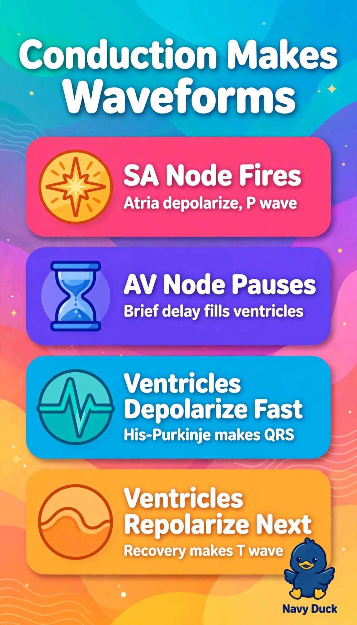
Short Explanation
Once you link each squiggle to its heartbeat job, strips stop looking scary. Conduction flows like a relay race handoff, so atria pass the baton to ventricles.
Full Explanation
Normal conduction begins at the sinoatrial node producing atrial depolarization seen as the P wave, pauses briefly at the atrioventricular node to allow ventricular filling, then travels rapidly through the bundle of His and Purkinje fibers producing ventricular depolarization seen as the QRS complex, followed by ventricular repolarization seen as the T wave. Stating the AV node fires first to make QRS and the SA node fires ventricles to make P reverses pacemaker hierarchy and misassigns waveforms to chambers. Stating the SA node fires ventricles to make QRS and Purkinje fibers make P misplaces impulse origin and conduction speed and mislabels repolarization sources. Stating ventricles fire first to make P and atria make QRS inverts the entire sequence and misrepresents normal upright T wave physiology. Exam caveat is to recognize normal order and shapes without diagnosing arrhythmias beyond PCT scope, and a practical check is to trace P to QRS to T in order and confirm one P before each QRS when reviewing rhythm.