
A cardiology technician reviews a telemetry strip for a stable adult with a regular rhythm, narrow QRS complexes, and one upright P wave before each QRS with consistent PR intervals. The nurse asks what the waveforms show about electrical activity. Which description of the cardiac conduction system and waveform components is accurate?
Select an answer to reveal the explanation.
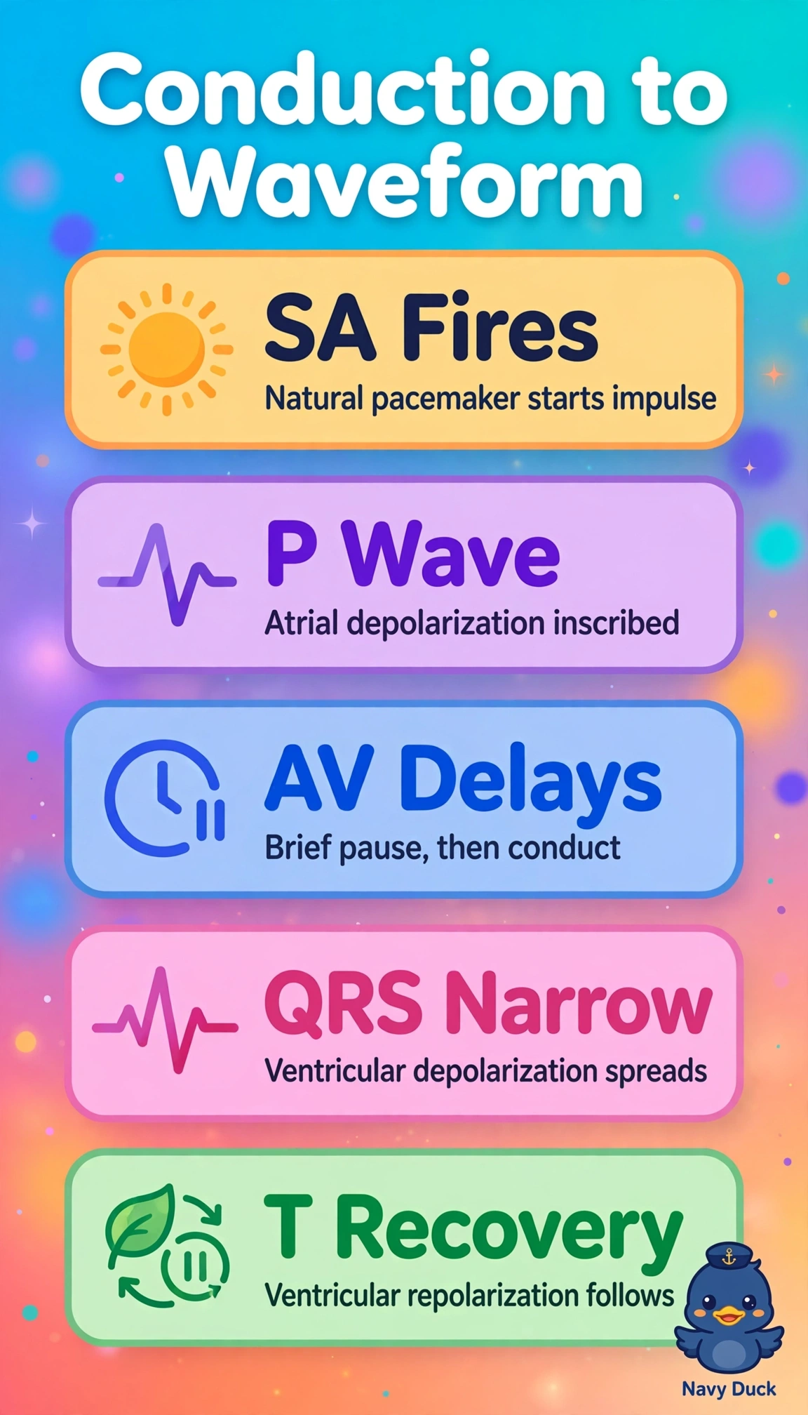
Short Explanation
You're watching a tidy relay where your SA node fires first and lights up the atria as the P wave. That spark then zips down like a baton pass to the ventricles to create the QRS.
Full Explanation
Normal conduction begins at the sinoatrial node, spreads through atria producing atrial depolarization inscribed as the P wave, delays at the atrioventricular node, then travels through bundle branches and Purkinje fibers to depolarize ventricles inscribed as the narrow QRS, with repolarization seen later as ST-T changes. Describing SA initiation, P as atrial depolarization, and QRS as ventricular depolarization captures sequence and waveform physiology. Attributing P to ventricular repolarization and QRS to atrial contraction reverses chamber and electrical-mechanical concepts because repolarization follows depolarization and QRS is ventricular electrical activity. Assigning T to atrial depolarization and QRS to filling confuses filling mechanics with electrical inscription because T represents ventricular repolarization and filling is mechanical diastole. Calling QRS ventricular repolarization and T atrial recovery inverts depolarization-repolarization order because QRS is ventricular depolarization and atrial repolarization is normally hidden. On the exam, link each deflection to depolarization or repolarization of a specific chamber, and in practice confirm one P per QRS and PR consistency for sinus rhythm.