
A technician in an outpatient clinic prepares a resting 12-lead EKG on a calm adult with a regular pulse, must verify normal sinus rhythm for the tracing, and recalls the sinoatrial node initiates each beat. The provider expects atrial depolarization, ventricular depolarization, and repolarization to appear in order. Which cardiac conduction-to-waveform sequence should the technician recognize to confirm the expected components?
Select an answer to reveal the explanation.
Short Explanation
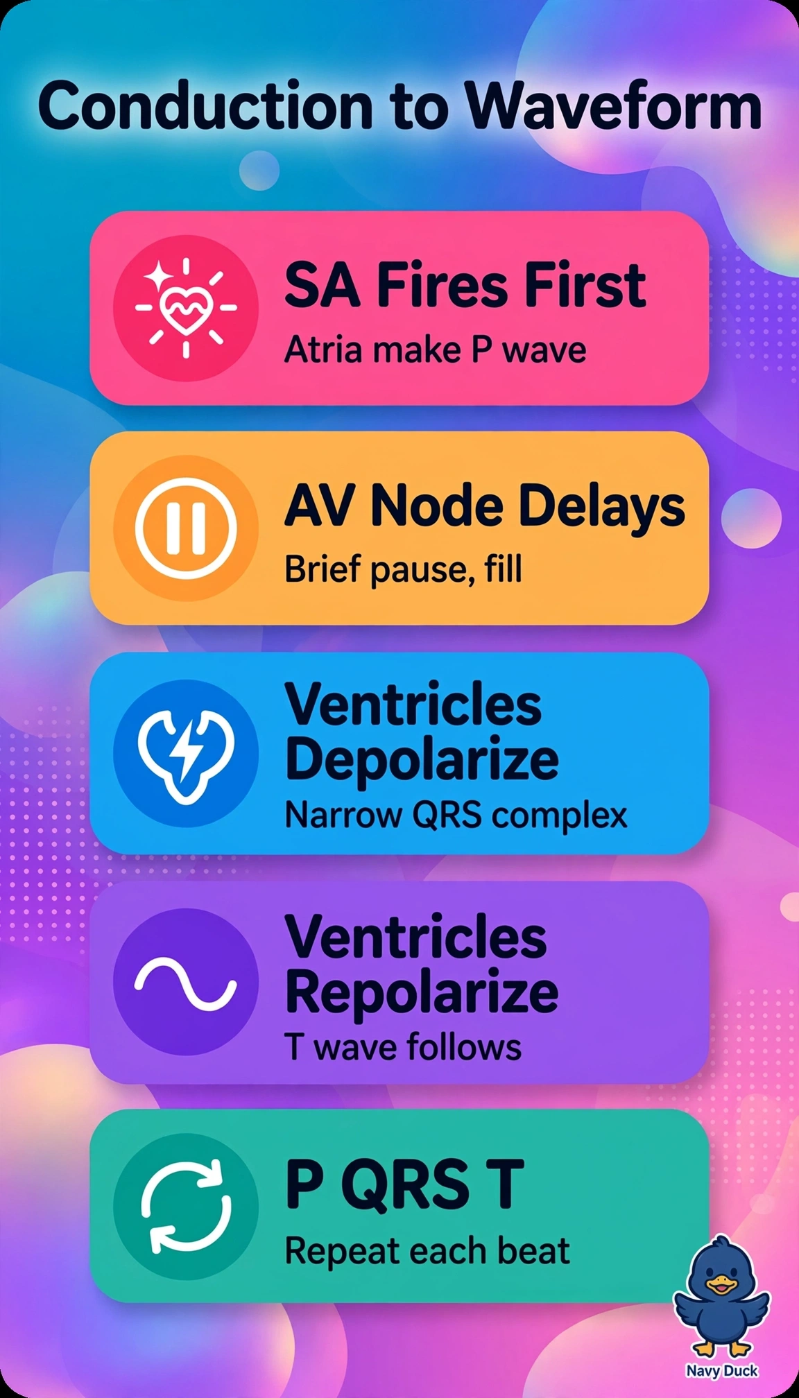
You can read rhythm like a relay race with a planned handoff. The SA-to-AV sequence links each P wave to a narrow QRS and T in order.
Full Explanation
Normal sinus rhythm originates in the sinoatrial node which depolarizes atria to inscribe a uniform P wave before each QRS, followed by physiologic slowing at the atrioventricular node creating the PR interval for ventricular filling, then rapid His-Purkinje conduction depolarizes ventricles to produce a narrow QRS with repolarization inscribing the T wave, so the correct sequence links SA initiation to P, AV delay, then QRS-T in required order. The sequence without AV delay fails on conduction physiology because loss of nodal slowing impairs filling and mimics accessory-pathway conduction, so immediate QRS after P cannot be accepted as normal even when order appears preserved. The sequence ending after QRS without a T wave fails on completeness because repolarization must be visible for QT, ischemia and electrolyte review, so required repolarization is unconfirmed. The sequence attributing the P wave to the AV node fails on pacemaker hierarchy because sinus P waves must arise directly from SA firing, while junctional activation yields absent, inverted or retrograde P waves indicating non-sinus rhythm despite preserved QRS-T. An exam caveat is that regular pulse alone does not prove sinus origin since ectopic rhythms can be regular, requiring P morphology and consistent P-to-QRS relation. A practical check is to verify an upright P before each narrow QRS with stable PR and a distinct T after each QRS in lead II.