
In a small urgent care room you must obtain a routine 12-lead EKG on an older adult who has a below-knee left leg amputation with a healed stump and a right modified mastectomy with a flat chest dressing, and the patient is anxious about lead placement. The machine allows quality tracings with documented modifications. Which EKG adaptation correctly balances accurate placement with respectful modification for amputation and mastectomy?
Select an answer to reveal the explanation.
Short Explanation
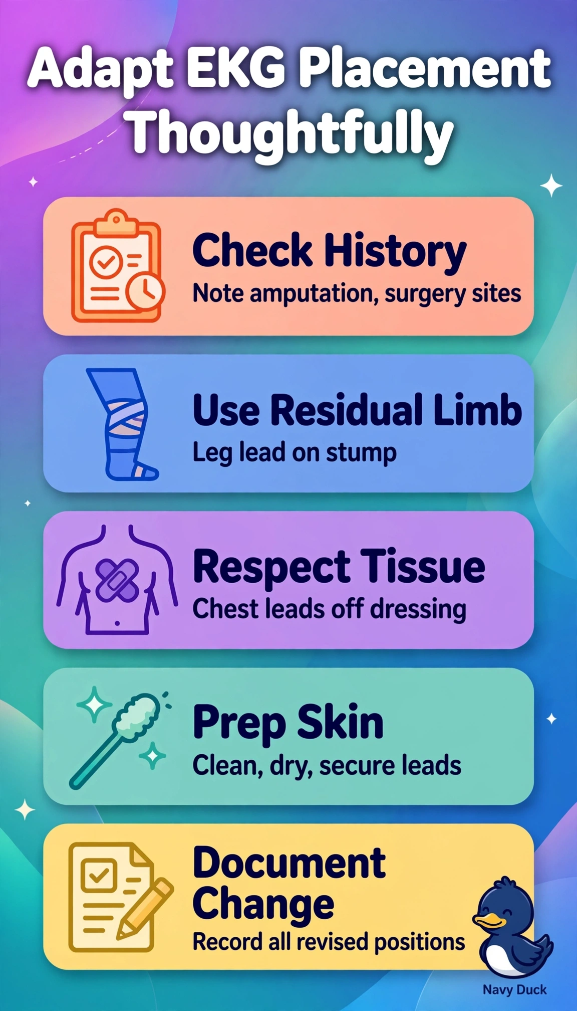
You're working with the body you've got today, not the textbook diagram. You're keeping continuity of the electrical picture by using the residual limb for the missing-limb lead and writing down exactly what you changed.
Full Explanation
The correct adaptation works because the electrode for an amputated limb belongs on the residual limb, as far distal as the healed skin allows, which keeps the lead closest to its standard limb position and preserves Einthoven’s triangle and frontal-plane vectors better than any torso site; precordial electrodes go on exposed skin at the closest available landmarks, off the flat post-mastectomy dressing, and explicit documentation lets the interpreter account for the changes while calm explanation addresses the patient’s anxiety. Moving the leg lead to the lower torso is the fallback only when no usable residual limb exists, and here a healed below-knee stump is available, so the torso site needlessly shifts the vector and changes QRS amplitude and axis. Placing chest electrodes over the dressing to preserve exact spacing fails on skin-electrode interface concept because fabric increases impedance, attenuates R-wave progression and invites baseline wander and displacement, so the closest exposed landmarks with notation of shifted positions is required. Gathering both leg electrodes together on the right torso for symmetry fails on vector concept because eliminating left-right separation collapses the inferior frontal plane, distorting leads II, III and aVF axis and voltage and mimicking limb reversal that confounds ischemia assessment. An exam caveat is that any shifted limb or precordial placement must be labeled as modified rather than routine because normal QRS amplitude and axis criteria assume standard limb positions. A practical check is to narrate each placement before touching, verify adhesion and a stable isoelectric baseline on the rhythm strip, and note the exact stump and chest deviations on the tracing and medical record.