
During EKG acquisition for a stable adult with palpitations, the tracing shows distinct P waves, narrow QRS complexes, and upright T waves with a regular rhythm. The nurse asks what electrical events the waveforms represent for documentation. Which description correctly links the cardiac conduction system to the EKG waveform components?
Select an answer to reveal the explanation.
Short Explanation
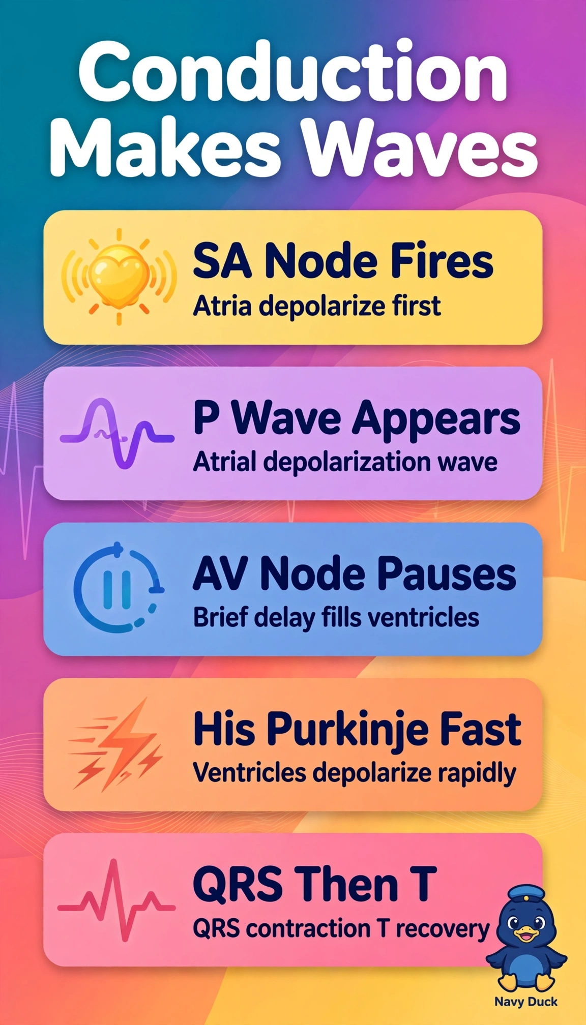
You're reading electricity in order from top to bottom of the heart. Atria fire first for P, then ventricles fire for QRS, which nails sequence every EKG depends on.
Full Explanation
Normal conduction begins at the sinoatrial node with atrial depolarization producing the P wave, followed by atrioventricular nodal delay that allows ventricular filling, then rapid His-Purkinje conduction producing ventricular depolarization inscribed as the QRS complex, with ventricular repolarization producing the T wave. The ventricular repolarization makes P description reverses chambers and phases, since P is atrial depolarization and the T wave reflects ventricular recovery, not atrial delay. The atrial repolarization makes QRS description misassigns both pacemaker roles, since QRS is ventricular depolarization and the ST segment represents early ventricular repolarization plateau, not SA activity. The bundle branches make P description inverts order and anatomy, since bundle branches activate ventricles for QRS while the P wave precedes them as atrial activity, not resting baseline. Exam caveat is to pair each waveform with both location and electrical direction, depolarization versus repolarization. Practical check is to confirm upright P before each narrow QRS when rhythm is sinus before troubleshooting artifact or lead placement.