
A 58-year-old woman has acute pleuritic chest pain, tachycardia, and hypoxemia after knee surgery. Chest radiograph shows a wedge-shaped peripheral opacity, ECG shows sinus tachycardia with nonspecific T-wave changes, and CT angiography is delayed. Which imaging interpretation best explains her presentation while awaiting definitive CT?
Select an answer to reveal the explanation.
Short Explanation
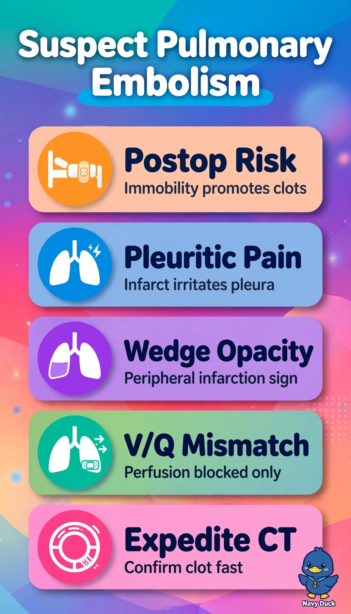
Clot pain after surgery plus a wedge infarct is telling you something. A V/Q gap explains hypoxemia without airway collapse, pointing to embolism first. Treat the clot until CT says otherwise.
Full Explanation
The picture favors acute pulmonary embolism pending CT angiography because knee surgery with immobilization confers high thrombotic risk and abrupt pleuritic pain with tachycardia and hypoxemia reflects ventilation-perfusion mismatch and pleural irritation from peripheral infarction, the wedge-shaped opacity representing Hampton hump rather than collapse or diffuse edema, while sinus tachycardia with nonspecific T changes is typical of acute strain and does not prove primary ischemia. Basal atelectasis from splinting soundly explains postoperative hypoxemia and tachycardia but fails radiographically because atelectasis yields linear plate-like opacities with volume loss and vascular crowding, not an isolated non-shrinking peripheral wedge with pleuritic pain. Demand ischemia after surgical stress soundly links tachycardia, T-wave changes, chest discomfort and hypoxemia but fails clinically because ischemic pain is pressure-like rather than pleuritic and provides no mechanism for wedge infarction, missing the key pleural-imaging fact. Postoperative pneumonia soundly links immobilization, pleuritic pain, hypoxemia and peripheral opacity but fails on acuity and context because bacterial consolidation typically evolves with fever, productive cough and air bronchograms rather than abrupt infarction immediately after orthopedic surgery. Exam caveat is that Hampton hump and S1Q3T3 are specific but insensitive, so absence never excludes embolism and D-dimer cannot rule out high-probability cases. Practical check is continuous monitoring, blood pressure for instability, Wells assessment, bleeding evaluation before empiric heparin, and expediting CT.