
An ICU nurse reports a water-seal chest drainage system shows continuous vigorous bubbling in the water-seal chamber, tidaling absent, and a portable SpO2 monitor reads 78% with good waveform while the patient is alert and pink with no distress. Suction is set at -20 cm H2O. Which troubleshooting sequence best resolves chest drain and pulse oximeter dysfunction?
Select an answer to reveal the explanation.
Short Explanation
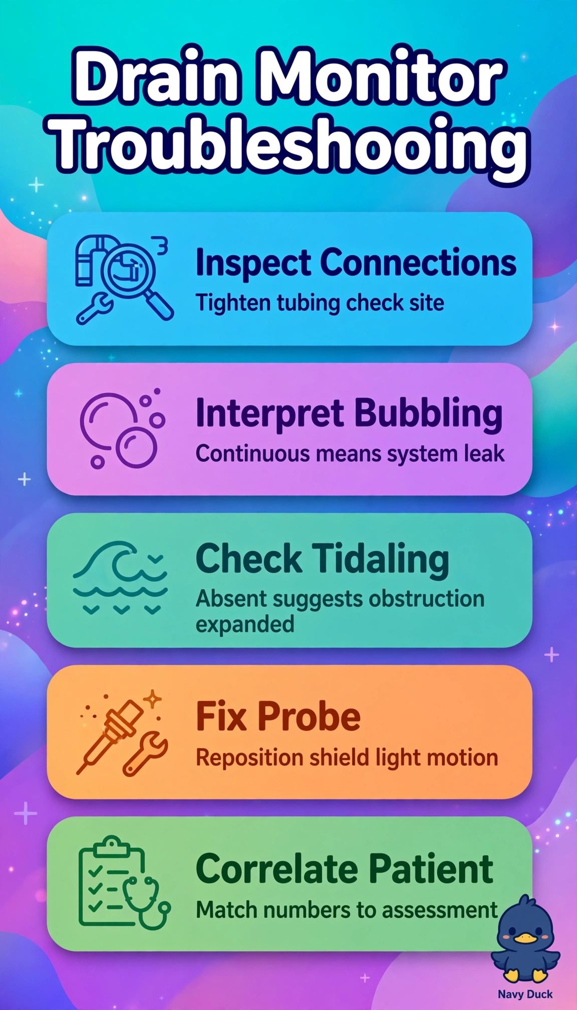
You can start by checking the chest-drain connections for an air leak and assessing probe placement, light interference, and perfusion. A discordant low SpO₂ in a comfortable, pink patient calls for checking the sensor before escalating care, while suction stays at -20 cm H₂O.
Full Explanation
The best sequence checks chest-drain connections for an air-leak source and assesses probe placement, light interference, and perfusion while keeping suction at -20 cm H₂O. Continuous vigorous bubbling in the water-seal chamber with absent tidaling suggests air entering the system through a leak, so inspect the connections; increasing suction will not seal the leak. A reading of 78% with a good waveform in an alert, pink patient without distress is discordant with the clinical picture, so check the oximeter before treating the number as true hypoxemia. Tightening connections and obtaining an arterial blood gas addresses a possible leak and could verify hypoxemia, but it skips the simpler bedside checks for probe placement, light, and perfusion before sampling and oxygen titration. Repositioning the probe and warming the extremity addresses possible oximeter error, but increasing wall suction to control bubbling is inappropriate: added negative pressure can amplify bubbling and does not correct an air leak. Examining connections and moving the sensor to another finger while maintaining suction addresses a leak and probe placement, but leaves light interference and perfusion unassessed. Exam caveat: do not increase suction to stop vigorous bubbling or act on an isolated oximeter value without clinical correlation. Practical check: inspect the connections and assess probe placement, light interference, and perfusion.