
A 46-year-old with six months of dyspnea on exertion has spirometry showing FVC 62% predicted with normal FEV1/FVC ratio, no bronchodilator response, and normal oximetry at rest. The physician suspects respiratory muscle weakness versus interstitial disease and orders complete pulmonary function and muscle strength assessment. Which pulmonary function testing sequence should the therapist perform to distinguish muscle weakness from parenchymal restriction?
Select an answer to reveal the explanation.
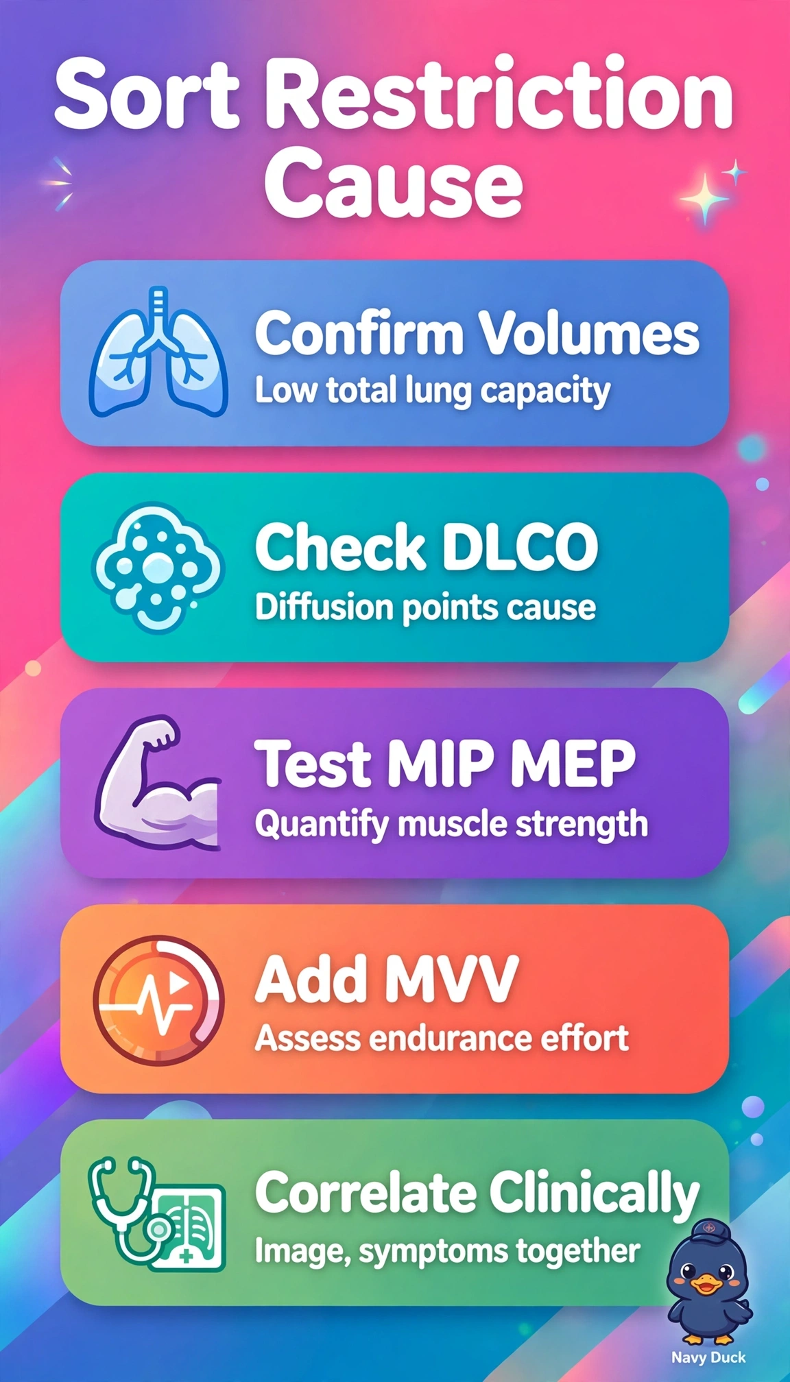
Short Explanation
You need more than one snapshot to solve this puzzle. Adding volumes, diffusion, and muscle pressures is like checking engine, tires, and battery together. That full set reveals restriction-versus-weakness patterns clearly.
Full Explanation
The low FVC with normal ratio suggests but does not prove true restriction, which requires low total lung capacity by plethysmography, and distinguishing interstitial disease from weakness requires both diffusion and strength data. DLCO helps separate parenchymal loss with reduced gas exchange from extraparenchymal weakness where DLCO corrected for alveolar volume is often preserved, while MIP from residual volume, MEP from total lung capacity, and 12-second MVV quantify inspiratory, expiratory, and endurance muscle performance that forced spirometry cannot. Plethysmographic volumes with DLCO alone accurately assesses parenchymal restriction and gas exchange but fails because without pressures and MVV neuromuscular weakness cannot be confirmed or excluded. Volumes with MIP, MEP, MVV and blood gases assesses mechanics and hypoventilation from weakness but fails because without DLCO interstitial fibrosis cannot be distinguished. Spirometry with volumes, DLCO, MIP and MEP but without MVV is incomplete because it omits endurance and sustained ventilatory capacity relevant to exertional dyspnea with normal resting oximetry. Exam caveat: low VC with normal TLC implies poor effort, air-trapping, or weakness, not fibrosis. Practical check: calibrate plethysmograph, coach maximal efforts, correct DLCO for hemoglobin, and add exercise oximetry after core testing.