
A 58-year-old former smoker with progressive dyspnea completes pulmonary function testing showing reduced FEV1/FVC ratio, increased total lung capacity and residual volume, and markedly reduced DLCO with normal hemoglobin. Which pattern interpretation should the therapist document for obstructive disease with diffusion impairment?
Select an answer to reveal the explanation.
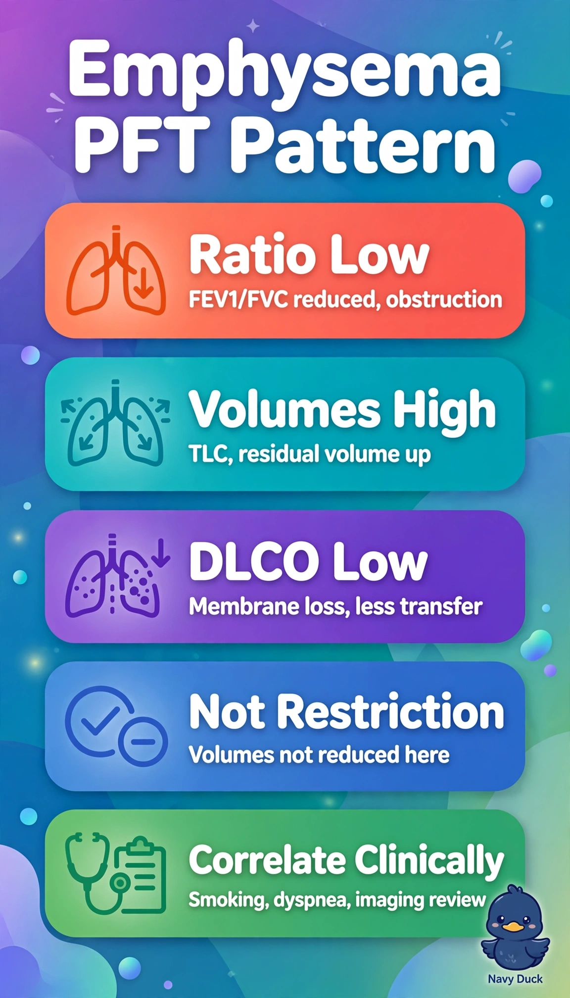
Short Explanation
You are reading footprints the lungs leave behind on paper. Low ratio says blocked, big lungs say trapped, low DLCO says walls destroyed. Emphysema ties all three clues together perfectly.
Full Explanation
The correct pattern fits emphysema because persistent airflow limitation with hyperinflation plus marked diffusion impairment after hemoglobin correction points to alveolar septal and capillary bed destruction rather than airways disease alone, matching progressive dyspnea in an older former smoker. The chronic bronchitic airway inflammation explanation fails conceptually because bronchitis produces obstruction with mucus hypersecretion and often hyperinflation but preserves alveolar surface so DLCO remains normal or high, and cannot account for marked reduction without parenchymal loss. The reversible asthmatic bronchospasm explanation fails because asthma is episodic and reversible with normal to increased DLCO due to intact alveoli and increased pulmonary blood volume, not progressive fixed obstruction with membrane loss in a smoking-exposed adult. The coexistent interstitial fibrosis explanation fails because fibrosis creates restrictive physiology with reduced TLC, RV and lung compliance, opposite the increased volumes seen here, while combined obstruction-restriction would lower volumes rather than cause hyperinflation. An exam caveat is not to equate any low DLCO with emphysema without checking hemoglobin correction, carboxyhemoglobin, alveolar volume, breath-hold time and spirometric quality. A practical check is to confirm hemoglobin-corrected DLCO and VA, review flow-volume loops and lung volumes for repeatability, correlate CT emphysematous changes, and document smoking pack-years, dyspnea course and bronchodilator response before finalizing.