
A 62-year-old with sudden dyspnea, tachycardia, hypotension, jugular venous distension, clear lungs, right ventricular strain on ECG, and swollen tender calf after long travel develops worsening hypoxemia. Which management priority best addresses suspected pulmonary embolism with shock?
Select an answer to reveal the explanation.
Short Explanation
You paired clot thinking with circulation support instead of waiting. Stabilizing pressure while starting anticoagulation workup protects the right heart, like bracing a bridge while engineers inspect it.
Full Explanation
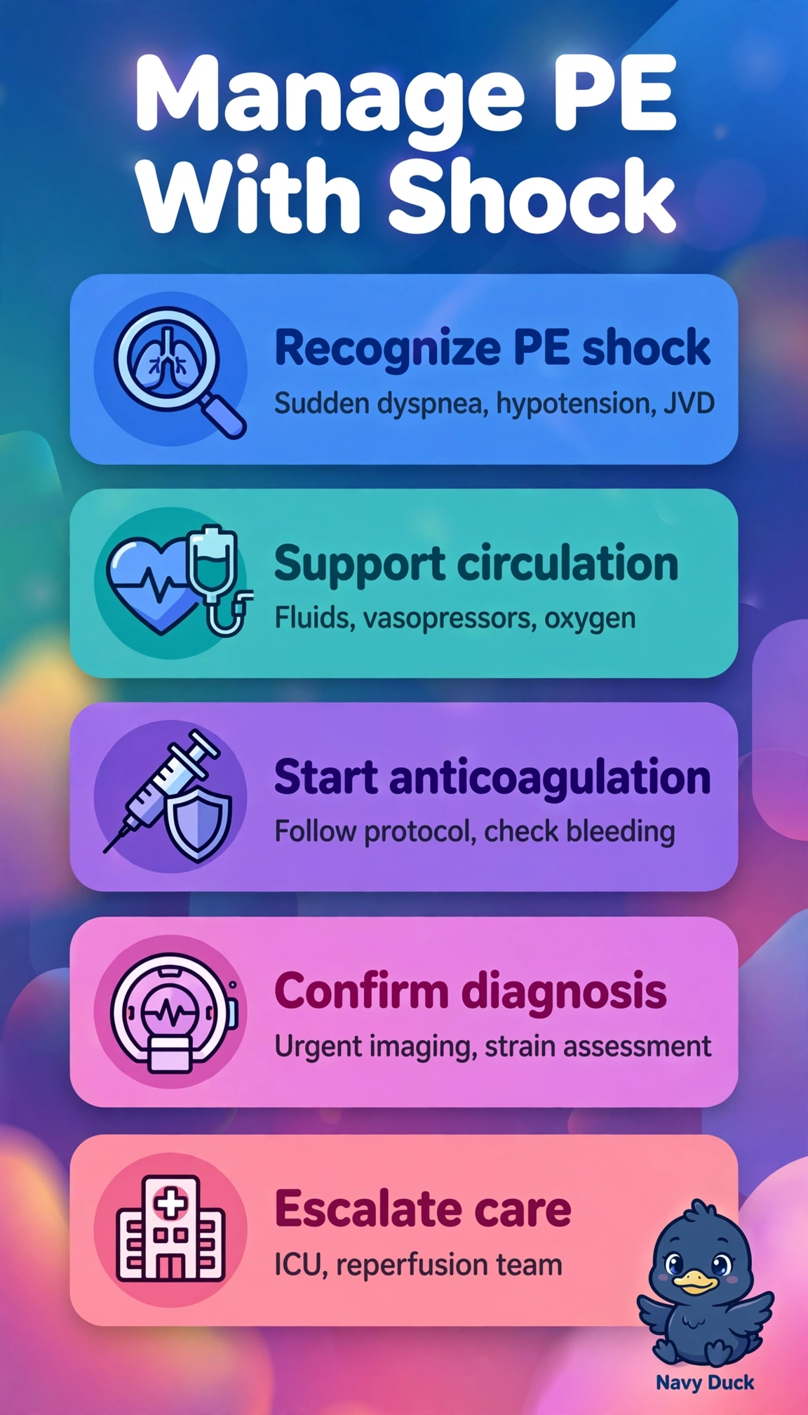
This presentation of sudden dyspnea, tachycardia, hypotension, jugular venous distension, clear lungs, right ventricular strain, tender swollen calf after long travel and worsening hypoxemia strongly suggests high-risk pulmonary embolism causing obstructive shock from impaired right ventricular outflow and reduced left preload, so the priority is simultaneous perfusion support with cautious fluids and early norepinephrine, immediate therapeutic anticoagulation to halt clot propagation, and expedited definitive pulmonary arterial imaging to confirm burden and guide escalation to thrombolysis or embolectomy. The strategy using prophylactic-dose heparin despite appropriate fluids, norepinephrine and urgent CT angiography fails by intensity concept because prophylaxis does not treat established unstable thromboembolism and permits extension. The strategy confirming with leg ultrasound alone despite appropriate fluids, norepinephrine and therapeutic heparin fails by anatomic concept because calf thrombus proves venous thrombosis but does not define pulmonary arterial occlusion, burden, or alternative chest pathology. The strategy supporting shock with large-volume fluids and oxygen alone despite therapeutic heparin and urgent CT angiography fails by hemodynamic concept because persistent hypotension with right ventricular failure requires vasopressor support and excess fluids can worsen interdependence and output. Exam caveat is that clear lungs with JVD, shock and hypoxemia points toward obstructive physiology rather than sepsis or left failure and should not trigger D-dimer delays in instability. Practical check is to reassess pressure, lactate, oxygenation, right ventricular function, weight-based heparin dosing with bleeding risk, and CT readiness while preparing escalation if shock persists.