
A 66-year-old two days after hip surgery develops sudden pleuritic chest pain, tachycardia, tachypnea, hypoxemia, hypotension, jugular distension, and right heart strain on bedside ultrasound suggesting obstructive shock. Which cardiopulmonary management priority should the therapist implement for suspected pulmonary embolism with shock?
Select an answer to reveal the explanation.
Short Explanation
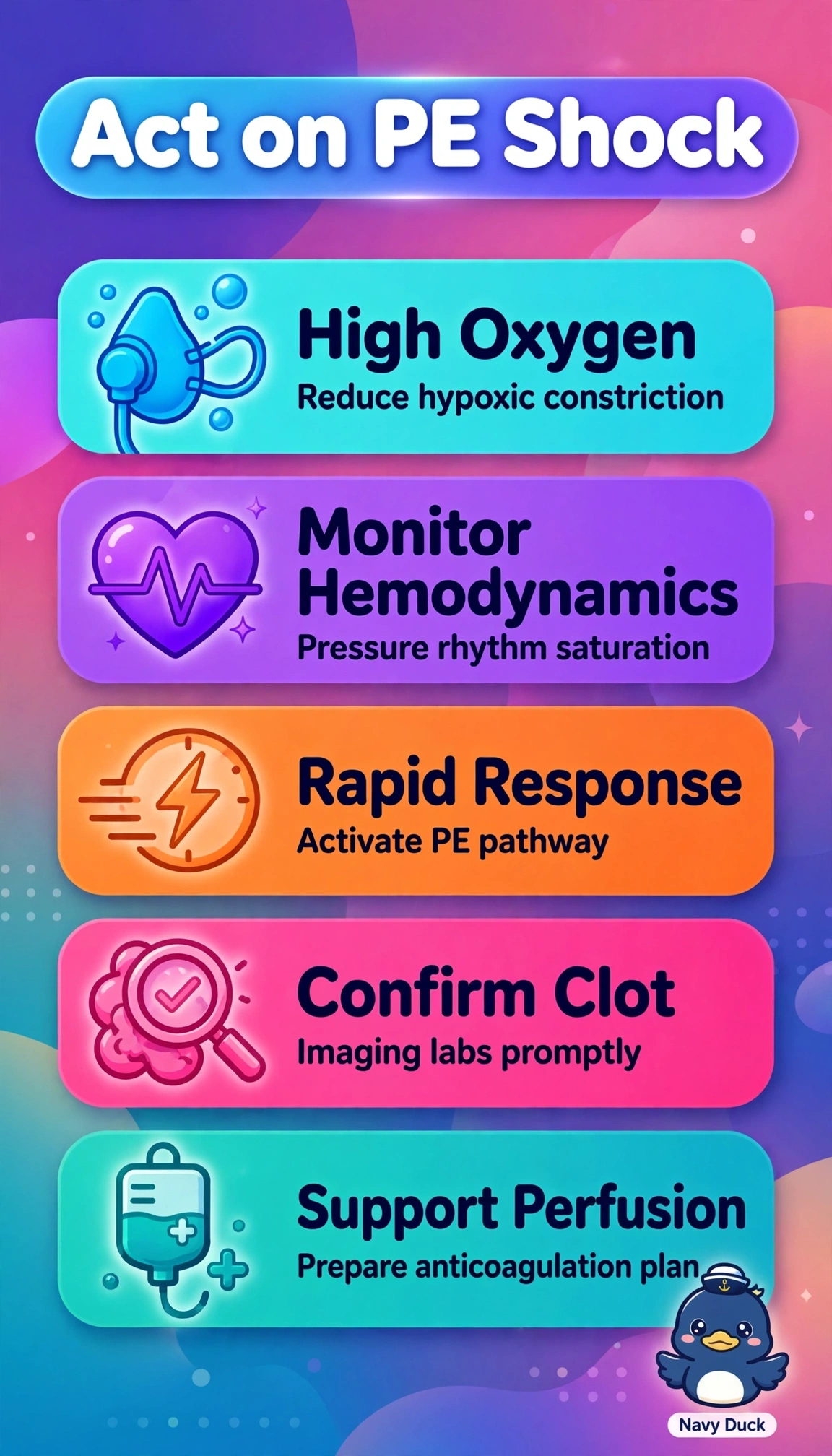
You’re dealing with a clogged pipe right when your lungs need flow most, so you’ll keep oxygen humming and eyes on vitals while help sprints to you. Don’t wait — get that rapid response going fast to protect circulation until the clot gets definitive care.
Full Explanation
Acute pleuritic pain, tachycardia, tachypnea, hypoxemia, hypotension, jugular distension and right heart strain two days after hip surgery indicate obstructive shock from suspected massive pulmonary embolism with impaired right ventricular output and left ventricular preload, so immediate high-flow oxygen for severe mismatch and work of breathing plus hemodynamic monitoring for pressure and perfusion trends with rapid response activation for definitive angiography and reperfusion evaluation is the priority. The low-flow conservative target strategy fails on oxygen delivery because it suits stable hypercapnia risk to limit hyperoxia but cannot meet high inspiratory demand and shunt in tachypneic shock and permits worsening hypoxia. The pulse oximetry surveillance strategy fails on circulatory monitoring because obstructive hypotension demands blood pressure, rhythm, mentation, urine and lactate tracking and oximetry alone misses progressive right ventricular failure and perfusion loss. The routine consultation imaging strategy fails on urgency because shock requires emergency escalation, not standard queuing, which delays ICU transfer, anticoagulation decisions and reperfusion eligibility. Exam caveat is that stable suspected embolism allows routine workup, while hypotension plus right strain defines instability requiring resuscitation and emergent activation. Practical check is to verify high-flow function and saturation, ensure large-bore access, attach cardiac and frequent pressure monitoring, and relay vitals, jugular findings and ultrasound results clearly to the response team.