
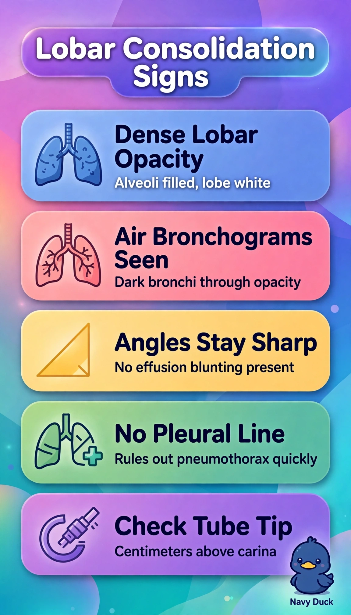
A 58-year-old mechanically ventilated patient develops fever, leukocytosis and increased purulent secretions. Portable AP chest radiograph shows dense left lower lobe opacity with visible air bronchograms, sharp costophrenic angles, no visceral pleural line, and endotracheal tube tip 4 cm above the carina. Which radiographic interpretation should the therapist document for this consolidation finding?
Select an answer to reveal the explanation.
Short Explanation
You've got all the clues pointing one way here. Air bronchograms inside a dense lobar opacity with sharp angles and no pleural line screams alveolar filling, so consolidation wins like footprints left in fresh snow.
Full Explanation
The correct documentation of left lower lobe consolidation with air bronchograms consistent with pneumonia and adequate tube position is supported because dense lobar opacity with patent air-filled bronchi silhouetted against fluid-filled alveoli defines consolidation, and with new fever, leukocytosis and purulent secretions in a ventilated patient strongly indicates ventilator-associated infection, while sharp costophrenic angles argue against large effusion, absence of visceral pleural line argues against pneumothorax, and endotracheal tip 4 cm above the carina is within the safe range requiring no manipulation. The right lower lobe pneumonia interpretation fails by laterality error because it correctly describes consolidation morphology but misidentifies side, misdirecting auscultation, suctioning, positioning and serial comparison. The mucus-plugging resorptive atelectasis interpretation fails by collapse-versus-filling confusion because preserved air bronchograms without fissural shift, volume loss or crowded vessels plus infection favor inflammatory alveolar filling needing antibiotics over airless collapsed lung needing clearance. The recommend-advancing-the-tube interpretation fails by airway mismanagement because 4 cm is adequate, not low, and unnecessary advancement risks mainstem intubation and lung injury. Exam caveat: portable AP films magnify structures and hide small effusions or apical pneumothoraces, so distinguish pneumonia with air bronchograms from atelectasis with volume loss, edema with bilateral bat-wing distribution, and effusion with blunted angles by integrating clinical signs. Practical check: measure tip-to-carina distance, verify bilateral breath sounds and pressures, and correlate film with temperature, white count, secretions and cultures before documenting.