
A 68-year-old ventilated patient in the ICU has stable SpO2 93-94% on FiO2 0.40, cuff pressure 24 cmH2O, flat capnography at 39 mmHg, but transcutaneous CO2 rising from 52 to 69 mmHg over 3 hours with increasing sedation and small tidal volumes. Which transcutaneous monitoring interpretation best confirms worsening hypoventilation?
Select an answer to reveal the explanation.
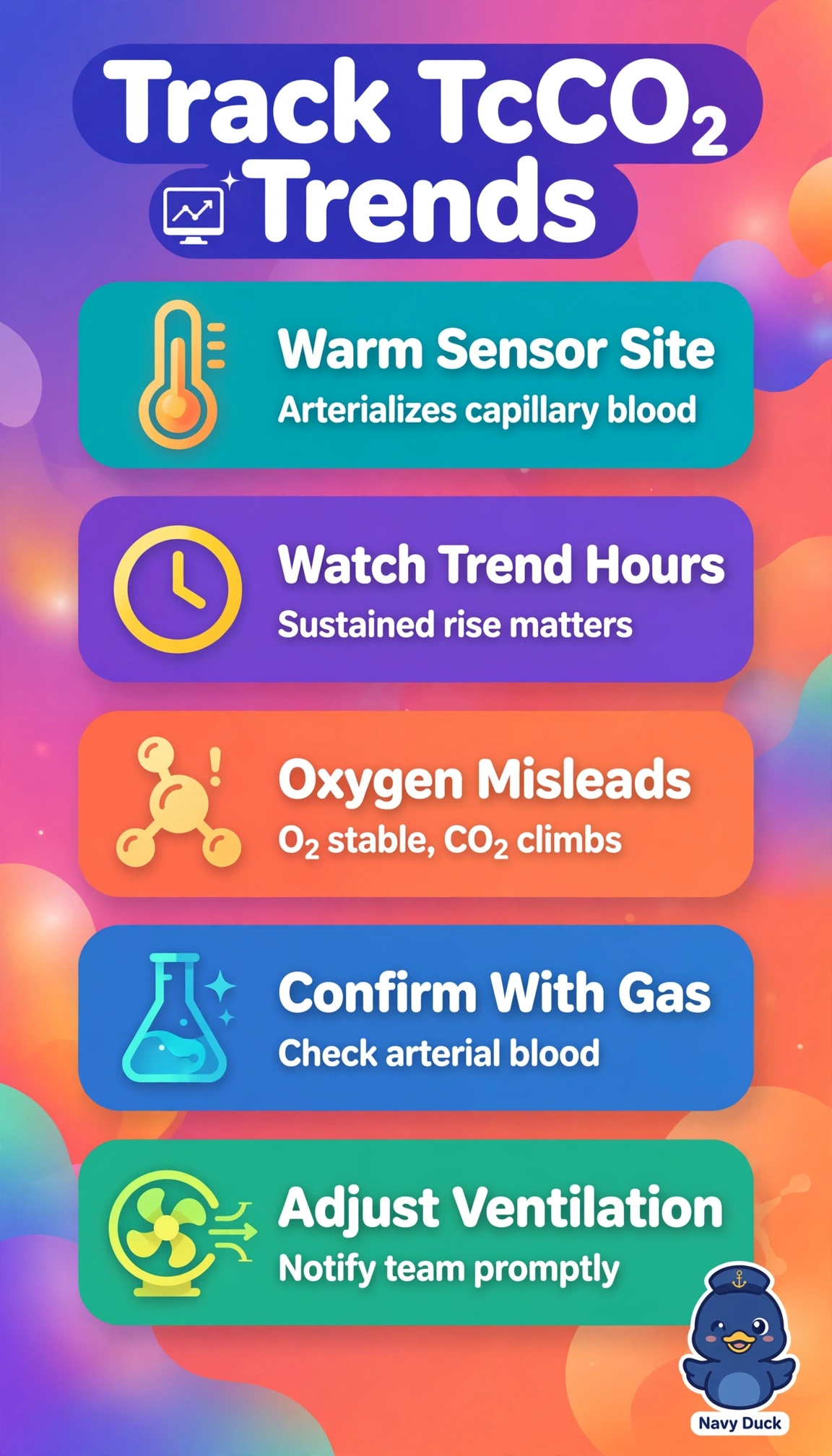
Short Explanation
You've got conflicting monitors, and you're right to feel torn. Steady oxygen doesn't mean ventilation's fine, so trust the trend like a slow-rising tide over single snapshots.
Full Explanation
The correct interpretation is true alveolar hypoventilation because a sustained transcutaneous rise from 52 to 69 mmHg over three hours accompanied by deeper sedation and small tidal volumes indicates falling alveolar minute ventilation, while stable saturation on FiO2 0.40 reflects preserved oxygenation and flat end-tidal 39 mmHg underestimates arterial CO2 when low exhaled volumes and increased dead space widen the arterial to end-tidal gradient. Raising inspired oxygen to wash out CO2 fails conceptually because oxygenation and ventilation are separate processes and added FiO2 does not increase CO2 elimination despite borderline oximetry. Recalibrating transcutaneous values to the capnography baseline fails because it assumes end-tidal CO2 equals alveolar and arterial CO2, which is unreliable with small tidal volumes and a flat waveform that dilute expired gas. Relocating the heated probe for suspected poor skin perfusion in an older patient addresses isolated artifact but fails because it disregards a prolonged directional trend plus clinical correlation and normal cuff pressure. An exam caveat is that normal oximetry and capnography never exclude hypercapnia on supplemental oxygen. A practical check is prompt arterial blood gas with review of exhaled minute ventilation, sedation depth, waveform, and probe placement and heating before adjusting ventilation.