
A 5-year-old with recurrent urinary infections is scheduled for voiding cystourethrography to assess reflux, needing bladder filling through a catheter, fluoroscopic imaging and voiding views. Which cystography technique best demonstrates vesicoureteral reflux and urethral anatomy?
Select an answer to reveal the explanation.
Short Explanation
You'll catch reflux best when the bladder's full and squeezing like a water balloon about to pop, so you've got to watch it fill and empty under fluoroscopy. That voiding cystourethrography approach lets you see urine climbing where it shouldn't, plus your urethral anatomy.
Full Explanation
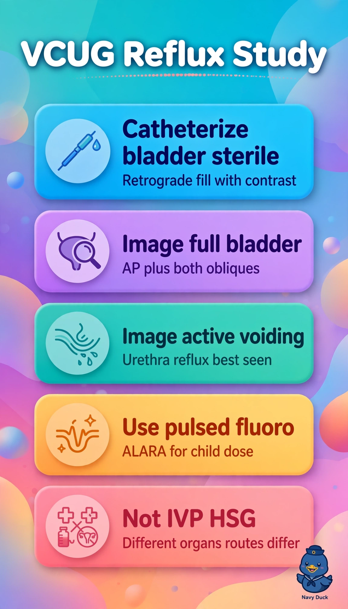
Direct fluoroscopic voiding cystourethrography is required because controlled retrograde filling through a catheter progressively distends the bladder and raises intravesical pressure to provoke vesicoureteral reflux while pulsed fluoroscopy captures filling, fully distended oblique and voiding phases, allowing grading of reflux, evaluation of bladder contour and ureterovesical junctions, and detailed urethral anatomy during active micturition, which together answer both clinical questions in recurrent infection; direct radionuclide cystography with intravesical tracer and gamma-camera monitoring is valid for low-dose reflux screening and follow-up and preserves catheter filling and voiding but fails because photon imaging lacks spatial resolution for posterior urethral valves and fine mucosal detail; CT cystography with dilute intravesical contrast is valid for traumatic bladder rupture and satisfies catheter distension but fails because static axial images without real-time fluoroscopy and a physiologic voiding urethral phase cannot show transient reflux or voiding anatomy; intravenous contrast with delayed voiding imaging is valid for upper-tract evaluation but fails because passive excretion without controlled retrograde distension does not reliably elicit or grade reflux; an exam caveat is that initial assessment with suspected anatomic abnormality demands fluoroscopic detail whereas radionuclide study suits surveillance of known reflux; a practical check is to verify sterile catheter placement, use weight-appropriate dilute contrast, use pulsed low-dose fluoroscopy with obliques, and document that voiding occurred under observation with postvoid views.