
A busy emergency portable chest shows no laterality marker, an unexpected dense circular shadow, and repeating fine lines across the anatomy. The charge nurse needs a repeat decision quickly. Which marker and artifact analysis best guides correct repeat action?
Select an answer to reveal the explanation.
Short Explanation
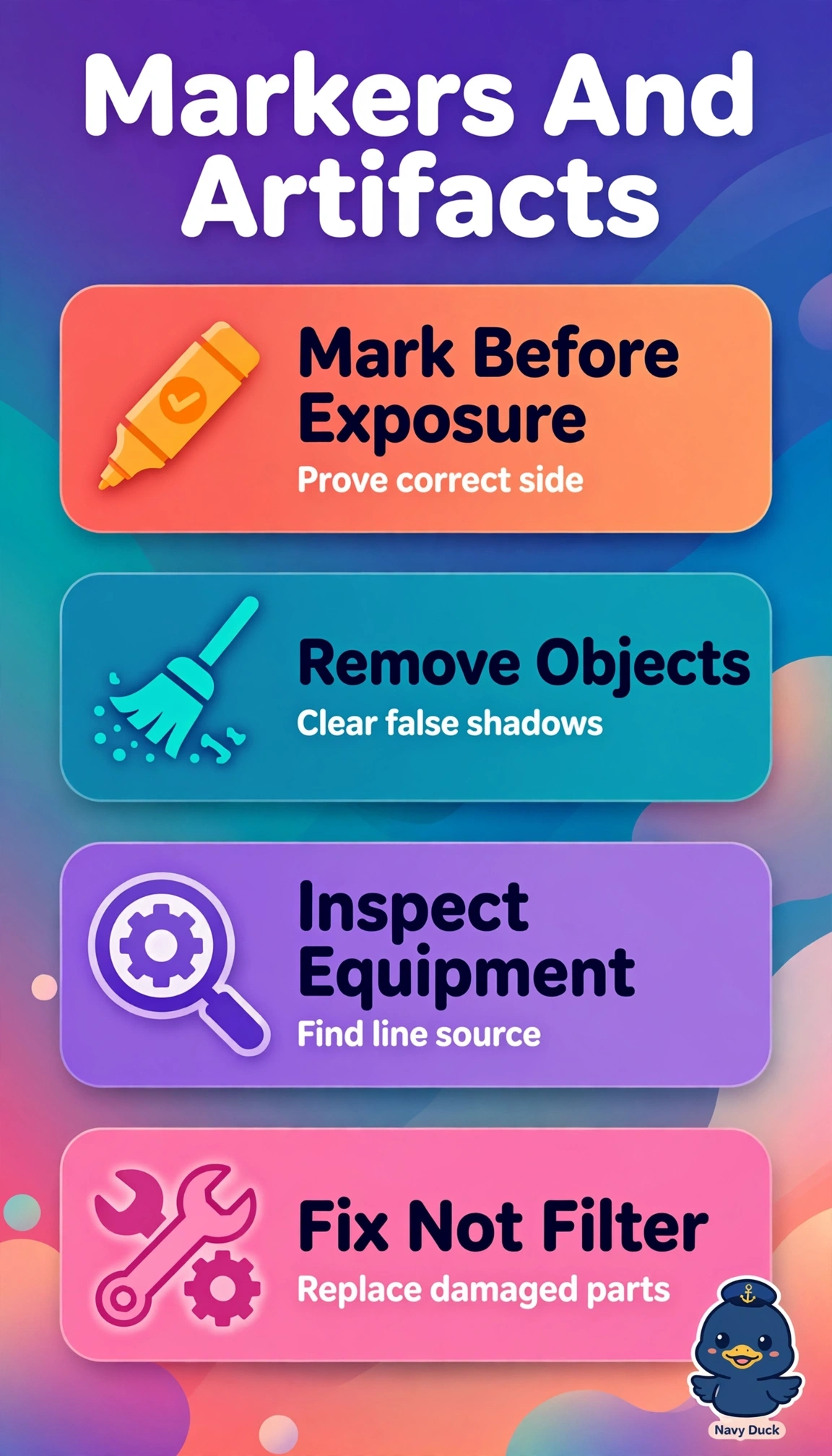
Sharp catch on three issues at once, and fixing each source beats patching the picture. Pre-exposure markers act like a signed label that proves which side you imaged.
Full Explanation
The correct triad restores diagnostic integrity because a physical anatomic marker placed pre-exposure provides legally defensible laterality that cannot be recreated afterward, systematic inspection identifies the unexpected dense circular opacity as removable external material such as an ECG snap, gown fastener or tubing rather than nodule or nipple shadow, and uniform repeating fine lines across anatomy point to cracked grid fibers or scored detector cover with persistent moire that technique changes cannot cure and therefore require equipment replacement before repeat. The post-exposure electronic annotation approach fails on marker legitimacy, since certification standards require the lead marker present during irradiation except narrowly documented exceptions, so correct object and grid handling still leaves laterality legally incomplete. Retaining the well-positioned ECG snap to save time fails on artifact versus anatomy, because any removable radiopacity in lung fields can mimic or obscure pathology and must be removed if clinically permitted rather than accepted for expediency. Re-angling and reprocessing to reduce moire fails on hardware fault resolution, because altered angle or filtering may soften pattern visibility but does not repair fractured grid that will recur. For examinations, distinguish regular geometric grid moire from random electronic noise or motion blur by its periodicity over unaffected anatomy. Before repeating, practically sweep eyes and hands over the chest, lift the gown, remove electrodes, inspect the cover and grid for cracks, and place the correct marker inside collimation without obscuring anatomy.