
A hypersthenic adult with a wide transverse abdomen and limited ability to hold a deep breath arrives for a supine KUB to check a ureteral stent, with a standard 35x43-cm detector available. The patient is comfortable supine but anatomy exceeds one field. Which adaptation should the technologist choose for body habitus and limited mobility during this KUB?
Select an answer to reveal the explanation.
Short Explanation
You're tailoring the fit, not forcing the patient to fit. Broad habitus needs wider coverage with overlap, like sewing two fabric panels for a full quilt.
Full Explanation
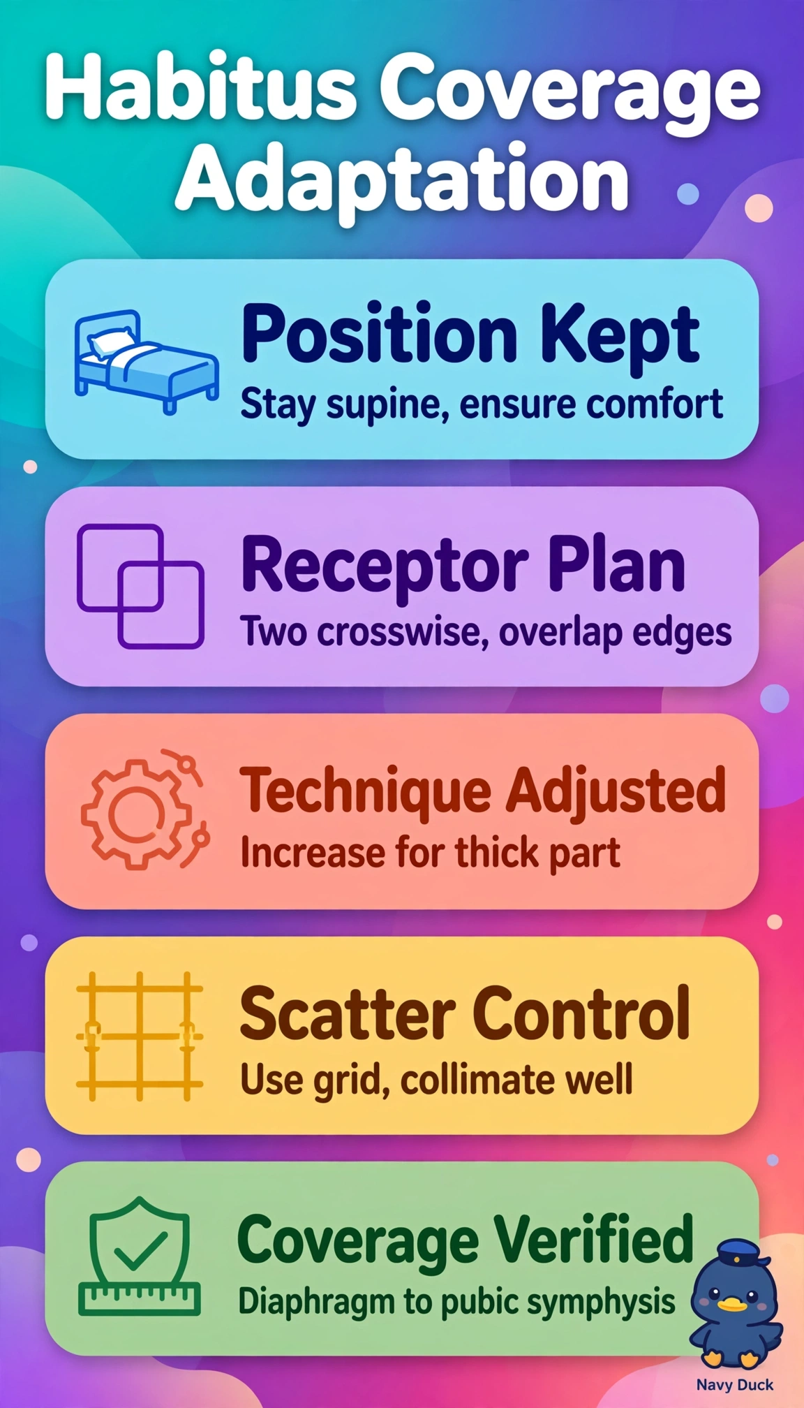
The correct adaptation succeeds because hypersthenic habitus is short, broad and thick, so placing the 35x43-cm detector crosswise maximizes transverse coverage, using two exposures with generous overlap avoids a gap or duplicated anatomy at the junction, including diaphragm to pubic symphysis in the supine position ensures the entire ureteral stent from renal pelvis to bladder is visualized, and modestly increased technique penetrates greater anteroposterior thickness to maintain density without excessive quantum mottle while shallow quiet breathing compensates for limited deep breath-holding. The crosswise two-field approach using standard technique fails by exposure concept because normal factors underpenetrate a thick hypersthenic abdomen and render the stent and soft tissues noisy and underexposed despite correct positioning and coverage. The lengthwise two-field approach with increased technique fails by orientation concept because portrait alignment suits tall asthenic patients and leaves lateral flanks clipped in a wide abdomen even with two fields and proper penetration. The crosswise two-field approach demanding deep inspiration fails by respiration concept because it requires a sustained deep hold the patient cannot perform, inviting motion, misregistration at the overlap and repeats, whereas short exposures during quiet breathing are more reliable. Exam caveat is to prioritize ordered supine stent coverage over erect air-fluid level techniques or dose reduction that omits anatomy or leaves tissue underpenetrated. Practical check is to confirm overlap by anatomic landmarks, verify superior diaphragm and inferior pubis on the preview, measure part thickness to set technique, and use immobilization and clear shallow-breathing instructions before exposing.