
A teenage athlete with a suspected scaphoid fracture and ulnar-sided wrist pain after a fall needs initial and specialized views without excessive rotation. The physician requests minimal displacement stress. Which wrist imaging combination best demonstrates scaphoid and ulnar-sided anatomy while limiting motion?
Select an answer to reveal the explanation.
Short Explanation
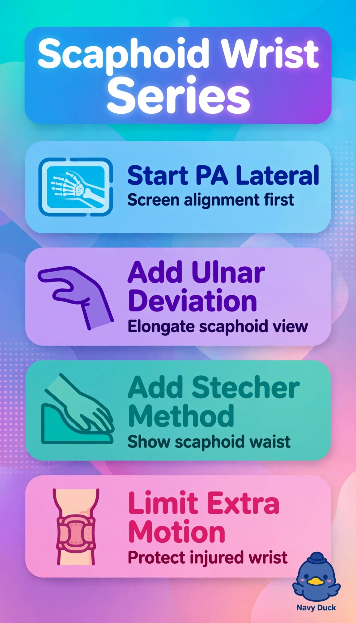
Smart positioning choice, because you stretched the scaphoid into view without stressing the injury. Ulnar deviation opens the scaphoid like pulling a telescope straight.
Full Explanation
Standard PA and lateral wrist establish alignment and general fracture screening, ulnar deviation elongates and profiles the scaphoid by reducing overlap, and the Stecher method with increased angulation further demonstrates the scaphoid waist where most fractures occur, together minimizing repositioning while evaluating ulnar-sided structures including triangular fibrocartilage region. The radial-deviated PA with Gaynor-Hart axial hand approach fails by positioning mechanism because radial deviation foreshortens and overlaps the scaphoid and Gaynor-Hart is a tangential sesamoid or carpal canal projection, not a scaphoid elongation view. The AP oblique hand with hyperflexed lateral and reversed canal approach fails by anatomy focus because hand obliques do not profile the wrist carpus and hyperflexion stresses injury without scaphoid elongation. The PA hand with pronated oblique elbow and tangential finger approach fails by region and projection because elbow and finger methods do not image carpal alignment or scaphoid waist detail. Exam caveat: distinguish Stecher scaphoid PA ulnar deviation from Gaynor-Hart tangential sesamoid and from Bridgeman or ulnar-deviation stress for instability. Practical check: keep wrist neutral except controlled ulnar deviation, angle as protocol directs, and immobilize to prevent motion.