
A 19-year-old collegiate wrestler reports jaw clicking and limited opening after years of strain, and the physician suspects temporomandibular joint dysfunction without acute fracture. The patient can open and close the mouth on command and can tolerate upright positioning with the head carefully aligned. Which TMJ projection strategy best demonstrates joint function in this patient?
Select an answer to reveal the explanation.
Short Explanation
You’ve got to catch the jaw in both gears to spot the hitch. Shooting open and closed side by side shows the glide at a glance, and translation is the motion that tells the story.
Full Explanation
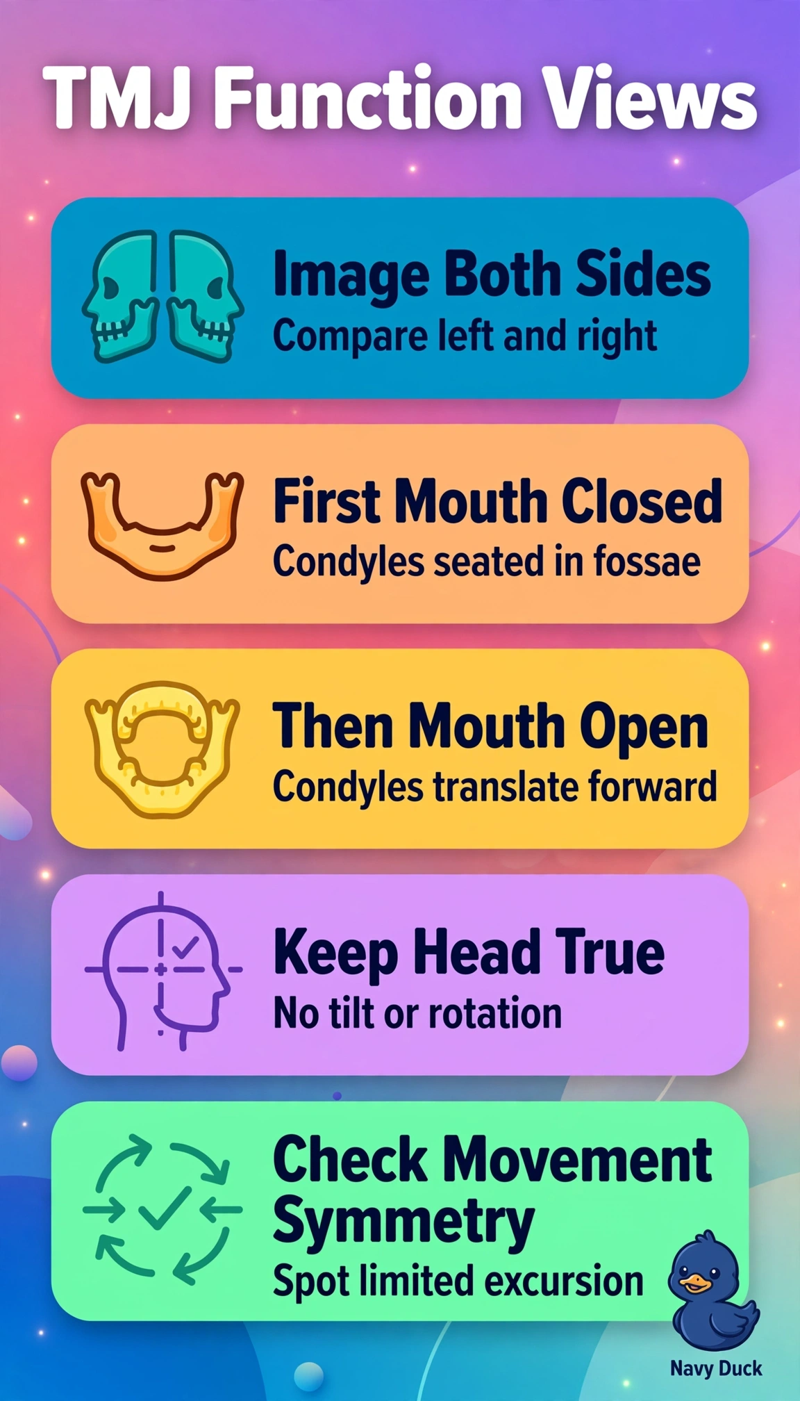
Correct strategy uses bilateral open- and closed-mouth projections because TMJ dysfunction is functional and requires comparison of condylar translation within each fossa from rest to maximal comfortable opening to demonstrate symmetry, excursion beneath the eminence, and limitation causing clicking in this cooperative patient without acute fracture who tolerates upright aligned positioning. Bilateral closed-mouth-only axiolateral imaging fails by function concept because it elegantly shows resting osseous relationships and joint spaces but cannot demonstrate dynamic anterior-inferior translation or restricted motion central to the clinical question. Unilateral open- and closed-mouth imaging of the painful side fails by comparison concept because it shows motion while appropriately considering dose but eliminates the contralateral control needed to recognize asymmetric hypomobility or hypermobility from chronic strain. Bilateral open- and closed-mouth imaging with head obliqued for comfort fails by alignment concept because rotation and tilt distort condyle-fossa relationships, alter joint spaces, and prevent valid side-to-side and open-to-closed comparison despite capturing motion. Exam caveat is to never force opening beyond tolerance and to halt functional views if acute fracture or dislocation is suspected in favor of trauma protocol. Practical check is to verify erect aligned head, use identical technique across all images, coach reproducible positions, and confirm symmetric forward condylar movement on open views.