
A repeat lateral lumbar spine image shows washed-out gray vertebrae, blurred trabeculae, elongated pedicles, and speckled noise despite standard positioning. The supervisor asks for a quality analysis before repeating. Which image evaluation analysis best separates contrast, resolution, distortion, and mottle causes?
Select an answer to reveal the explanation.
Short Explanation
You sorted four problems at once, and that systematic eye will save repeats. Image quality breaks down like a recipe where each wrong ingredient creates its own flavor.
Full Explanation
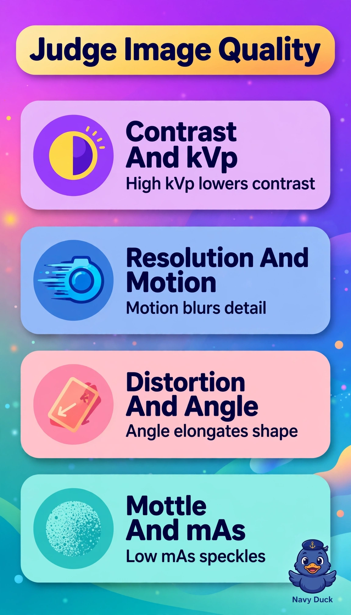
High kVp widens the exposure latitude and reduces differential absorption, producing low-contrast gray images, motion or large focal spot and low sampling blur fine trabecular spatial resolution, off-angle central ray or tilted anatomy elongates structures through distortion, and low mAs reduces photon quantity creating quantum mottle speckle that processing cannot fully smooth. The low kVp with high SID, grid cutoff, and high mAs interpretation fails by physical mechanism because low kVp increases contrast, high SID improves sharpness, grid cutoff causes underexposure lines rather than elongation, and high mAs reduces mottle. The short scale with small focal spot, detector size, and filtration interpretation fails by definition because short scale means high contrast, small focal spot improves resolution, detector size does not elongate, and filtration alone does not create photon-starved speckle. The collimation with low kVp, LUT, and tight collimation interpretation fails by causation because collimation affects scatter and field size, not intrinsic contrast loss, and lookup tables adjust display, not geometric elongation or quantum noise. Exam caveat: link each artifact to its primary exposure or geometry control to select the correct repeat correction. Practical check: assess histogram, edge sharpness, shape accuracy, and noise separately before adjusting technique.