
An intensive care patient needs bedside verification of a newly placed nasogastric tube for medication delivery, and a separate oncology patient needs PICC tip confirmation under C-arm fluoroscopy. The team requests low-dose real-time guidance. Which line placement imaging role best ensures safe PICC and enteric tube positioning?
Select an answer to reveal the explanation.
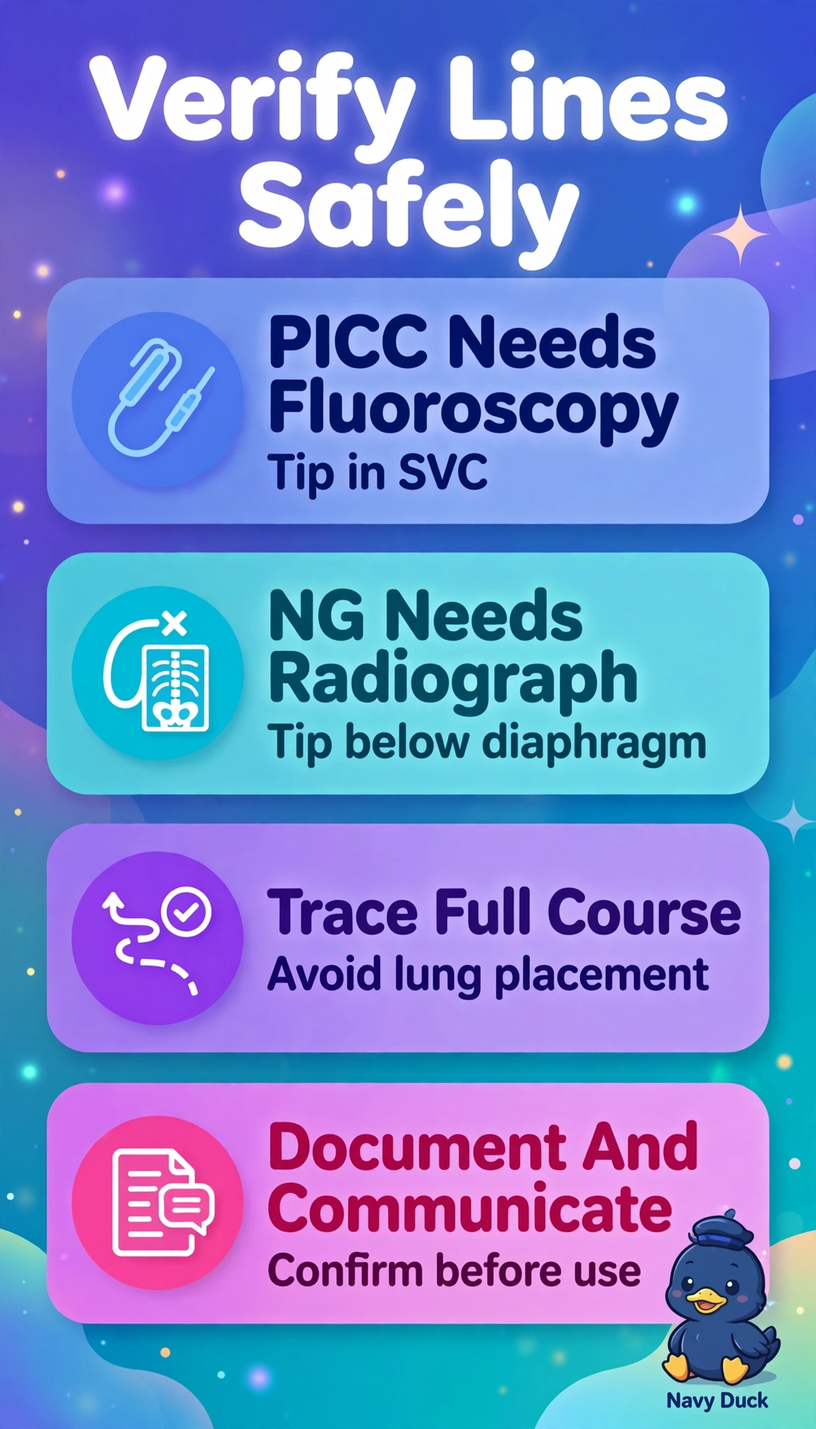
Short Explanation
You are keeping both lines safe by matching each tube to its right check. Tip location matters like parking in the right spot, not just anywhere on the street.
Full Explanation
PICC lines require tip termination in the superior vena cava or cavoatrial junction to allow high-volume infusion and prevent thrombosis or extravasation, best confirmed with real-time C-arm fluoroscopy during placement, while nasogastric or enteric tubes require radiographic proof that the tip and side ports pass below the diaphragm into stomach or specified small bowel to avoid pulmonary instillation before feeding or medication. The fluoroscopy-advance NG to duodenum with peripheral PICC approach fails by placement standard because NG tubes for medication usually target stomach and PICC tips must be central, not peripheral. The radiography-reposition PICC with fluoroscopy-inflate balloon approach fails by role scope because radiographers do not reposition central catheters sterilely and balloon inflation is not verified by fluoroscopy alone. The ultrasound-alone approach fails by modality limit because ultrasound cannot reliably trace NG course through aerated lung and bowel or document final PICC tip position for the record. Exam caveat: never approve feeding through an NG tube without documented below-diaphragm tip and side-port visualization on image. Practical check: trace the entire tube course, label right versus left, and communicate critical malposition immediately.