
A preschool child arrives with sudden choking, wheezing, and suspected peanut aspiration, now stable and cooperative. The physician wants to detect air trapping from a radiolucent airway foreign body. Which soft tissue and chest approach should the technologist select to evaluate movable airway obstruction?
Select an answer to reveal the explanation.
Short Explanation
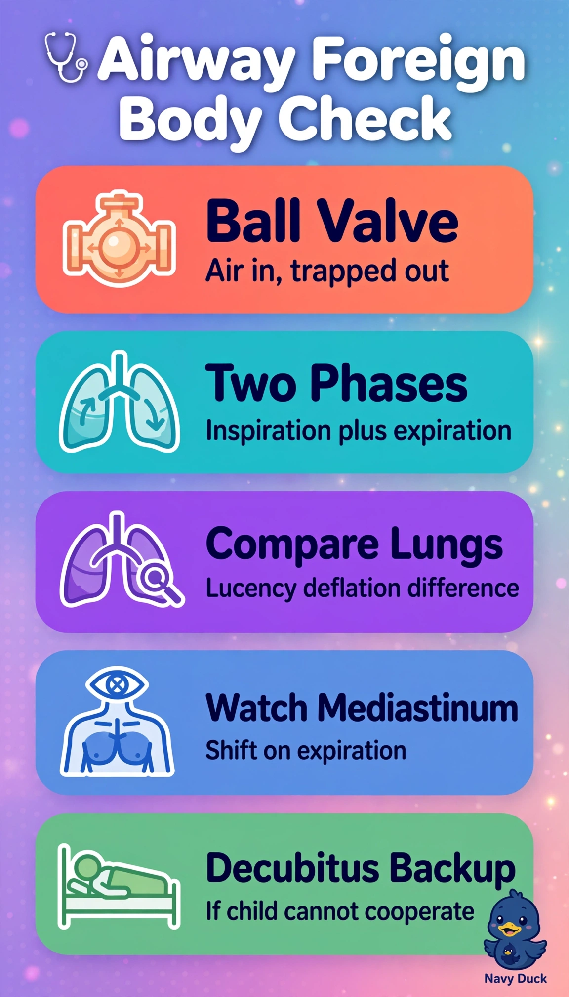
You can catch a sneaky peanut by watching how air moves, not by relying on one snapshot. Paired breathing phases are your before-and-after view of trapped air and mediastinal shift.
Full Explanation
Peanut fragments are radiolucent and rarely seen directly, so you look for ball-valve physiology: the affected lung may inflate on inspiration but trap air and remain relatively hyperlucent on expiration, with mediastinal shift toward the normal side. Paired inspiratory and expiratory PA chest images best demonstrate these changes in a stable, cooperative child; the PA projection supports symmetric comparison of lung aeration and mediastinal position. Erect inspiratory PA and lateral chest images are useful for other chest findings, but without an expiratory image they cannot demonstrate air trapping, and the lateral view does not show the frontal mediastinal shift. Paired inspiratory and expiratory lateral chest images can show diaphragm motion, but superimposed lungs limit side-to-side comparison of aeration and obscure frontal mediastinal movement. Inspiratory and expiratory lateral neck soft-tissue images can assess upper-airway narrowing, but wheezing after peanut aspiration suggests lower-airway involvement, and neck images cannot evaluate lung air trapping. An exam caveat is to distinguish upper-airway symptoms such as stridor, which may call for neck imaging, from wheezing that calls for chest evaluation. Practically, check for adequate inspiration (about eight to nine posterior ribs above the diaphragm) and expiration (crowding of the ribs and higher diaphragms), with no rotation and clear breathing instructions.