
A fluoroscopy suite upgrade includes variable collimation, added aluminum filtration, and positive beam limitation on the overhead tube. During acceptance testing the physicist asks about dose reduction compliance. Which beam restriction and filtration principle best satisfies NCRP 102 safety requirements?
Select an answer to reveal the explanation.
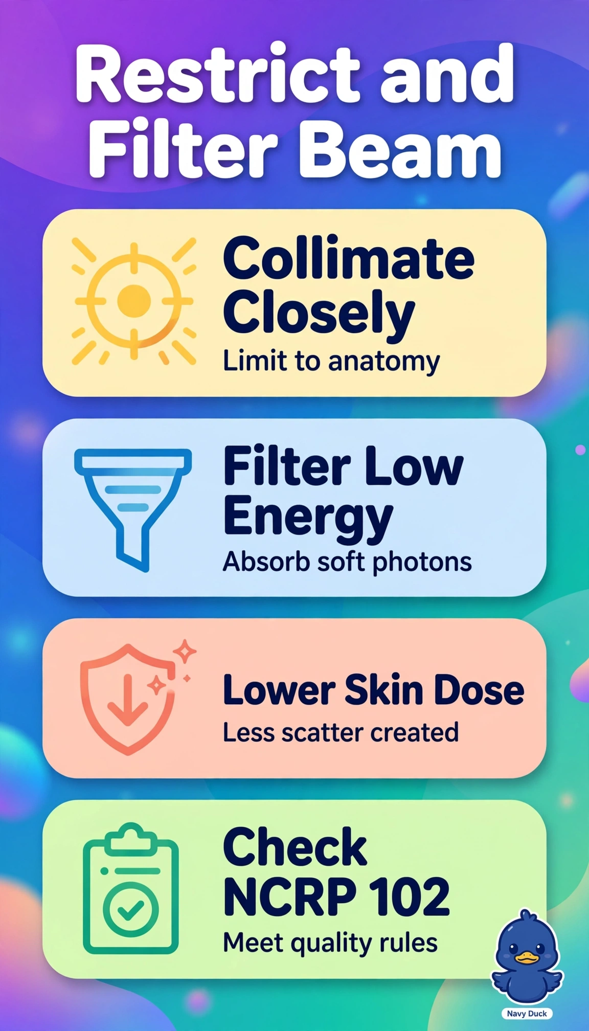
Short Explanation
Great job tightening the beam, because smaller fields and cleaner beams protect patients daily. Collimation works like window blinds that let light only where you need it.
Full Explanation
The correct principle combines the smallest field covering the anatomy with aluminum filtration meeting NCRP 102 half-value layer requirements because tight collimation lowers integral dose and scatter while filtration removes low-energy photons that add skin dose without forming the image. Collimating to the receptor with positive beam limitation and adequate filtration is routine practice but fails here because a receptor-sized field still irradiates tissue beyond the anatomy that tight coning would exclude. Tight collimation with filtration for improved contrast is familiar language but fails by concept because compliance hinges on absorbing low-energy photons to reduce entrance dose while maintaining half-value layer, not on hardening to sharpen contrast. Using overhead-tube positive beam limitation for the fluoroscopy field is tempting after that upgrade but fails by application because that system governs the overhead radiographic tube, while fluoroscopy needs its own tower-controlled variable collimation. An exam caveat is that writers contrast automatic receptor-sized limitation with manual anatomy-sized coning and expect the smaller anatomy-limited field plus verified filtration as dose-optimal. A practical acceptance check is to confirm half-value layer aluminum equivalence, verify light and radiation field congruence, and compare exposure for receptor-sized versus tightly coned fields.