
A 58-year-old outpatient with chronic right knee pain is scheduled for fluoroscopic contrast knee arthrography, can lie supine with the knee supported, reports no contrast allergy, and the radiologist needs sterile joint access with confirmed intra-articular placement before injection. Which arthrography workflow best ensures safe sterile access and placement?
Select an answer to reveal the explanation.
Short Explanation
You're keeping the joint clean and proving you're inside before you fill it. Sterile prep plus live confirmation delivers placement-assurance confidence like checking the door is open before carrying in groceries.
Full Explanation
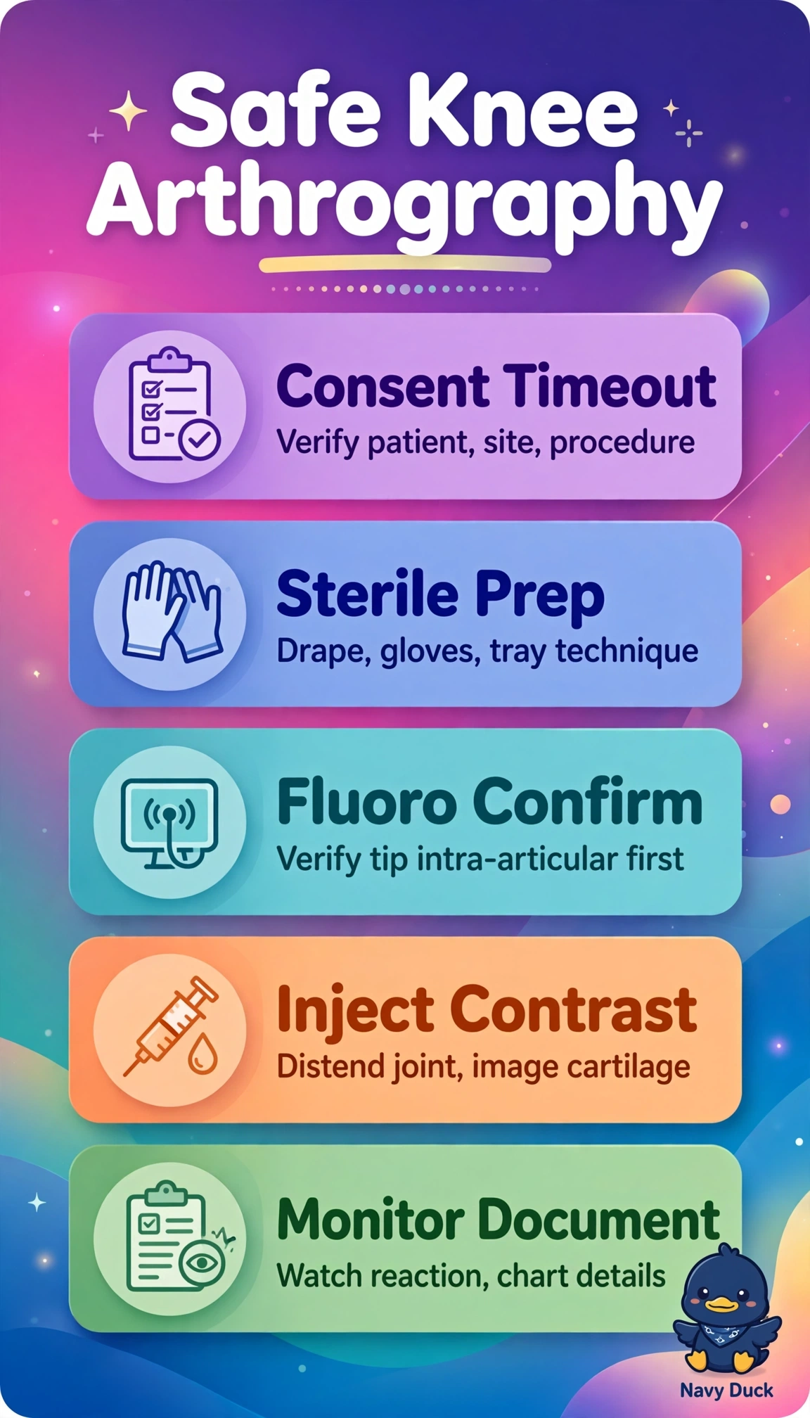
The correct workflow fits the ordered fluoroscopic diagnostic study because informed consent and timeout with right-knee verification establish safety and laterality, sterile prep and technique prevent septic arthritis, supine positioning with support allows stable access, and fluoroscopic confirmation before any substantive injection prevents extra-articular infiltration and ensures diagnostic distension in a patient without iodinated contrast allergy. Ultrasound-guided verification with iodinated contrast fails by guidance concept because although ultrasound is routine and excellent for knee injections, it cannot provide fluoroscopic bony-landmark confirmation and radiographic documentation of intra-articular contrast flow required for fluoroscopic arthrography. Fluoroscopically verified dilute gadolinium fails by contrast-physics concept because dilute gadolinium placed under fluoroscopy is standard for magnetic resonance arthrography, yet it lacks iodine radiopacity needed to opacify meniscal recesses and cartilage surfaces on fluoroscopy. Fluoroscopically verified corticosteroid without diagnostic contrast fails by purpose concept because image-confirmed steroid injection is standard therapy for chronic knee pain, but without iodinated contrast it gives no diagnostic distension or anatomic map and does not fulfill arthrography. Exam caveat is to distinguish guidance modality from diagnostic modality, since fluoroscopy may guide placement for magnetic resonance imaging without making the study a fluoroscopic arthrogram. Practical check is to reverify marking, allergy status, sterile tray and fluoroscopy function, then confirm free intra-articular flow with a tiny contrast test without resistance or soft-tissue blush before completing injection.