
A portable chest image from the intensive care unit looks very noisy with an exposure indicator far above the target range, yet anatomy is visible. The technologist used a much lower mAs than usual with a high kVp and full processing. Which exposure indicator interpretation best explains this gross exposure error?
Select an answer to reveal the explanation.
Short Explanation
Your detective work pays off because the number tells the exposure story behind the noise. Exposure indicator rises or falls inversely with detector dose, like a golf score dropping when you hit harder.
Full Explanation
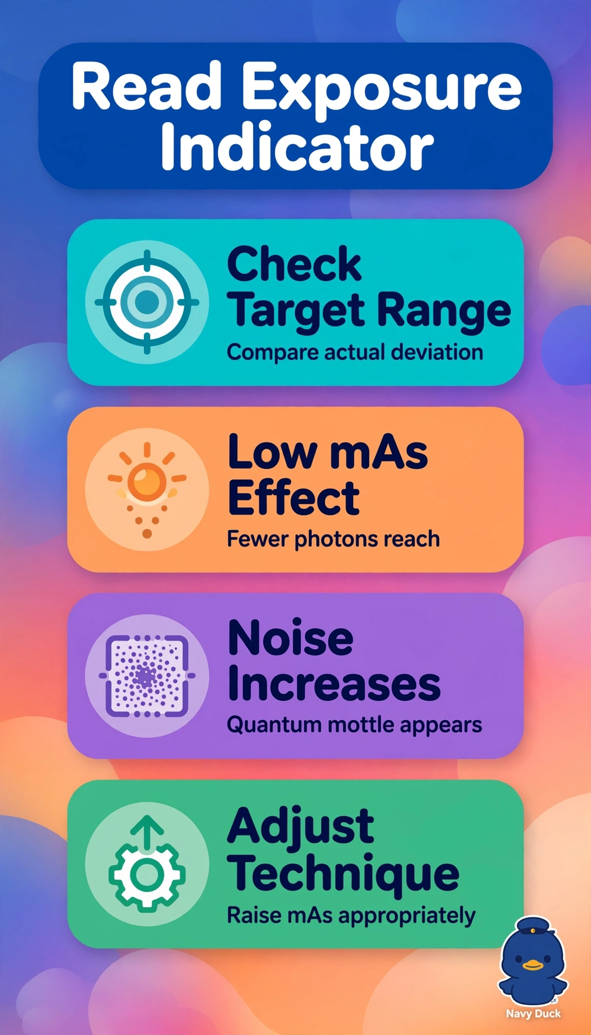
The correct interpretation recognizes severe photon starvation from much lower mAs as the driver of both findings because detector signal and quantum noise are governed primarily by mAs, while high kVp increases penetration and scatter but does not replace missing primary photons, and with full processing applied the system rescales brightness and contrast so anatomy remains visible yet pronounced mottle persists and the inverse-type exposure indicator rises far above target to flag underexposure requiring repeat. The overexposure-from-high-kVp reasoning fails by concept because true overexposure saturates the detector, lowers noise and drives an inverse-scale indicator downward, not upward with coarse graininess, and it overweights kVp while ignoring the stated large mAs reduction that dominates detector dose. Accepting the image because brightness looks normalized and anatomy is seen fails by concept because postprocessing normalization masks but never corrects quantum statistics, so visible anatomy does not equal diagnostic quality when deviation is gross and noise obscures subtle ICU lines and infiltrates. Blaming partial or failed processing fails by concept because the stem explicitly states full processing was applied and processing controls grayscale mapping, not photon count or indicator calculation, so it cannot explain coincident far-high deviation plus severe mottle. Exam caveat is to verify manufacturer scale direction before answering, since some indicators rise with dose and others fall, and brightness alone never confirms exposure. Practical check is to compare exposure index deviation to the target range, inspect the histogram and noise, then repeat with restored mAs per portable technique chart rather than compensating with extra kVp or reprocessing.