
A 6-year-old child is referred from pediatric endocrinology for short stature with an early growth spurt and no history of hand trauma. The order requests bone age assessment using minimal exposure and a standardized comparison method unaffected by dominant-hand use. Which projection and comparison standard should the radiographer use for bone age assessment?
Select an answer to reveal the explanation.
Short Explanation
You've got a growth question, not an injury puzzle, so you'll stick to the standard playbook. A single PA left hand-wrist keeps dose low and matches the atlas like checking height marks on a doorframe.
Full Explanation
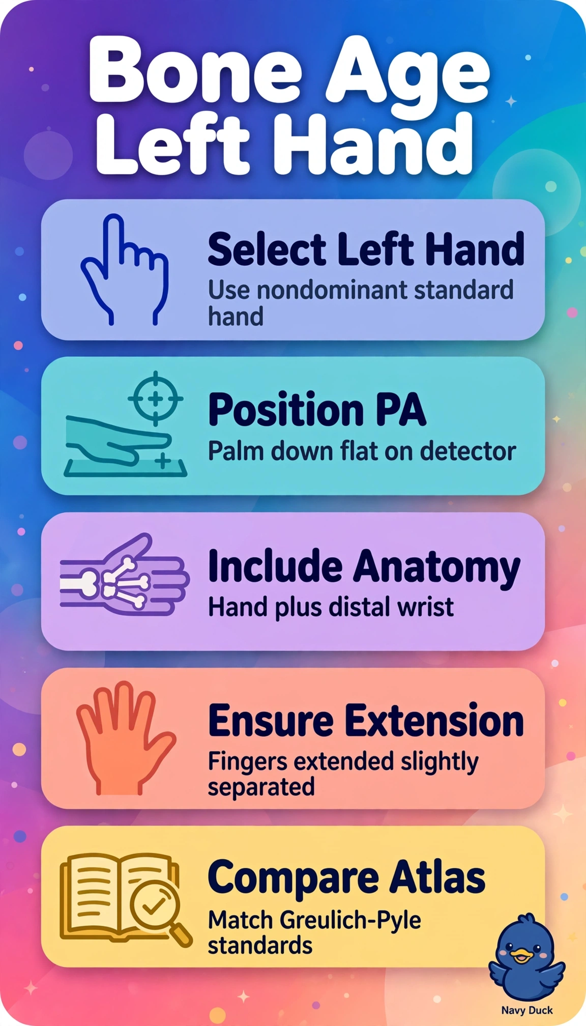
Bone age assessment evaluates ossification centers of the left hand and wrist against age-matched Greulich-Pyle atlas standards, because epiphyseal appearance and fusion follow a predictable sequence and the nondominant left hand avoids overuse changes that could skew maturation scoring. A single PA projection with the palm flat on the detector provides sharp bony detail with minimal object-to-detector distance and magnification, satisfying pediatric dose restraint while preserving atlas comparability. The right dominant-hand approach fails by laterality standardization, since atlas norms were validated on the left hand and dominant-hand stress can alter appearance; the AP projection approach fails by positioning geometry, since beam entry from the palmar side increases magnification and reduces sharpness compared with the standard PA method; and the bilateral-hands approach fails by dose optimization, since routine bilateral comparison adds unnecessary exposure without improving atlas matching when no trauma or asymmetry is suspected. For the exam, remember bone age is left hand-wrist PA for atlas comparison, while long-bone measurement and skeletal survey use entirely different multi-exposure protocols, and in practice verify left-hand markers, include the distal radius and ulna and all carpals, and confirm no jewelry or flexion obscures the epiphyses.