
Overnight portable AP chest on an intubated adult in ICU shows the medial clavicle ends asymmetric to the spine, the right lung apex clipped, and the scapulae overlapping the lateral lungs with the central ray angled low. Which positioning accuracy and tube-part-receptor alignment error does the image demonstrate?
Select an answer to reveal the explanation.
Short Explanation
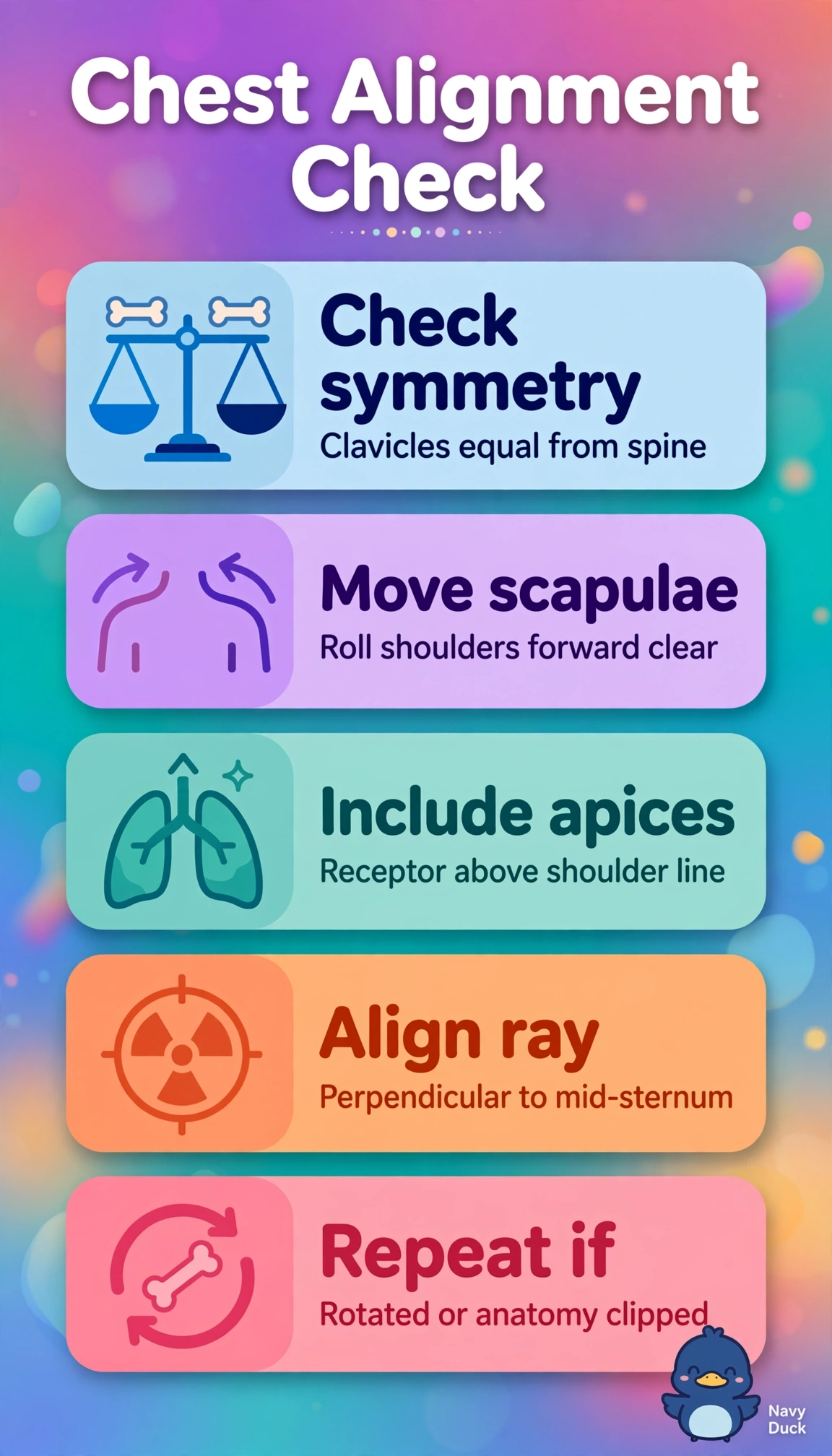
You nailed the clues if you linked asymmetry to rotation and clipping to alignment. Your tube-part-receptor geometry has to center all three together, like framing a photo squarely.
Full Explanation
Correct because asymmetric medial clavicle-to-spine distances define rotation, low downward ray depresses clavicles over apices and leaves scapulae over lateral lungs, and isolated right apical clipping with bases included proves receptor placed too inferior without supra-apical margin. The cephalad description fails tube-angle concept because superior angulation elevates clavicles above apices in lordotic projection and elongates anterior ribs rather than driving shadows inferiorly with scapular overlap. The tilted description fails motion concept because superior-inferior tilt alters vertical clavicle and rib levels, whereas unequal horizontal medial clavicle-to-spine distances specifically indicate left-right rotation about the long axis. The too-high description fails receptor concept because superior placement preserves apices and truncates costophrenic angles and bases, opposite to the stated apical loss that requires inferior placement. Exam caveat is supine portable ICU images magnify anterior structures and exaggerate rotation and angulation, so do not mistake scapular overlap or clavicular superimposition for inspiration or exposure failure. Practical check is center spinous process between clavicles, keep tube perpendicular unless angled view ordered, place receptor 1-2 inches above shoulders with arms rolled forward, and confirm apices and angles in light field.