
A portable chest image of a ventilated ICU patient looks grainy and mottled at a low mAs technique, while bony detail washes out on the workstation despite window adjustments. The detector and bit depth are unchanged. Which quantum noise, SNR, DQE, dynamic range, and bit depth principle best guides correction?
Select an answer to reveal the explanation.
Short Explanation
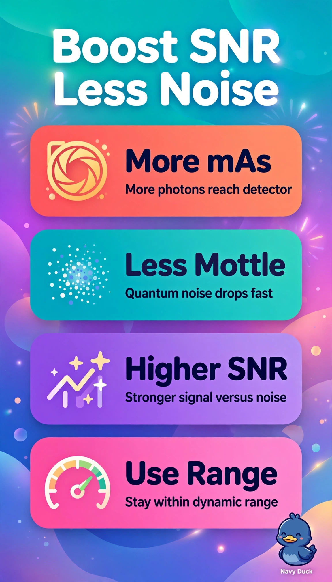
You're starving a photo of light and seeing static. Feeding it more photons smooths the grain with photon-sufficiency.
Full Explanation
The grainy mottled appearance at low mAs reflects photon starvation where too few quanta reach the detector, so quantum noise dominates and signal-to-noise ratio falls with the square root of detected photons, and the correct principle restores adequate statistics by increasing mAs while staying within detector dynamic range so unchanged detective quantum efficiency and bit depth can still encode sufficient gray scale for bone and soft tissue; the kVp-based approach fails by concept because although higher kVp increases transmission and can lower apparent mottle, it systematically reduces subject contrast and differential attenuation, which further washes out subtle bony detail already poorly visualized; the display-mapping approach fails by concept because window width and level only remap acquired pixel values to luminance without adding photons or improving intrinsic signal-to-noise ratio, so it cannot create missing information when adjustments are already exhausted with fixed detector and bit depth; the processing-based approach fails by concept because smoothing and frequency enhancement only change the presentation of noise and edges while true quantum signal-to-noise ratio remains limited by the original exposure, often trading suppression for loss of fine trabecular sharpness; for examinations remember mAs governs photon quantity and quantum noise while kVp governs photon quality, penetration and subject contrast, and do not confuse detector dynamic range and bit depth with display windowing; as a practical check confirm a modest mAs increase raises the exposure indicator without saturation, then reassess raw graininess and cortical visibility before further display or filtering refinement.