
A quality control technologist tests a 2-year-old mobile radiographic unit in the emergency department using a beam-alignment test tool and radiopaque markers, the light field looks centered, but prior images show unexplained collimation cutoff on one side. Service history is unremarkable. Which quality control evaluation should be performed to verify light field to radiation field congruence and central ray alignment?
Select an answer to reveal the explanation.
Short Explanation
Your eyes see light, but film sees X-rays. Congruence testing proves those two fields actually match where it counts.
Full Explanation
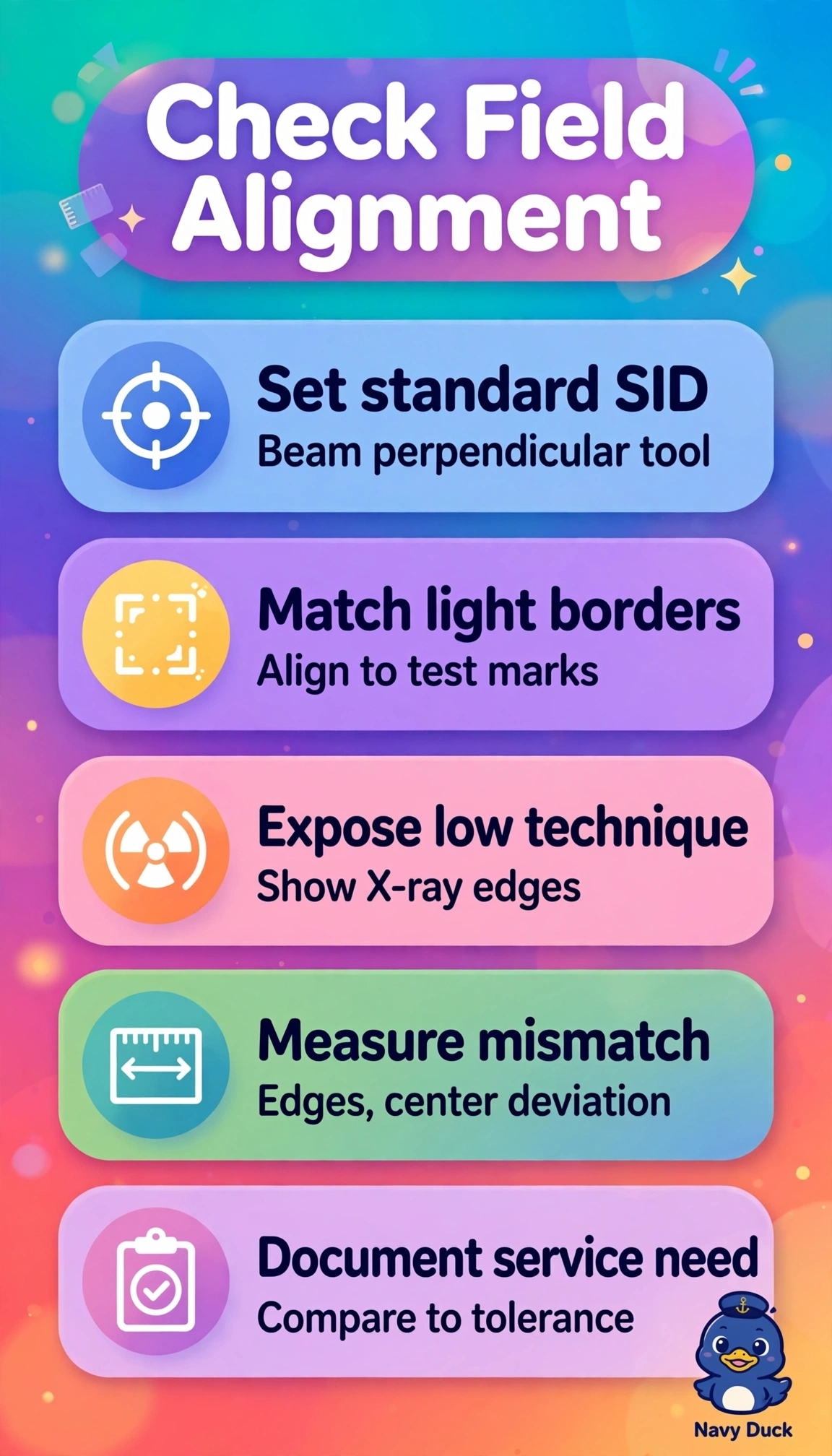
Light field to radiation field congruence and central ray alignment are geometric accuracy tests performed with a dedicated test tool containing radiopaque edges and a central ball, exposing under controlled SID to visualize the X-ray field on the image relative to the light field borders and to quantify perpendicularity, because misalignment causes unintended anatomy cutoff, repeat exposures, or excessive irradiated tissue even when the light looks centered. Exposing the tool and measuring edge mismatch and central deviation against department tolerance determines pass or fail and need for service. Widening collimation to hide cutoff and raising mAs masks the fault while increasing patient dose and scatter and fails to diagnose misalignment. Calibrating the exposure chamber and replacing the grid address exposure termination and scatter cleanup, not geometric congruence, so they would not explain unilateral cutoff. Measuring half-value layer evaluates beam quality and filtration with aluminum attenuators, relevant to kVp and patient dose but unrelated to field alignment. Exam caveat is to test at standard SID with the beam perpendicular and document results, and practical check is to align light borders to the tool markings, expose at low technique, and measure each edge discrepancy before calling service.