
A surgeon requests imaging for a middle-aged patient on postoperative day two after cholecystectomy with a T-tube in place, normal vitals, and concern for a retained common bile duct stone. Which imaging approach should the radiographer prepare to evaluate the biliary ducts through the surgical drain?
Select an answer to reveal the explanation.
Short Explanation
You're using the drain that's already there. T-tube injection gives a direct roadmap of the bile ducts without endoscopy.
Full Explanation
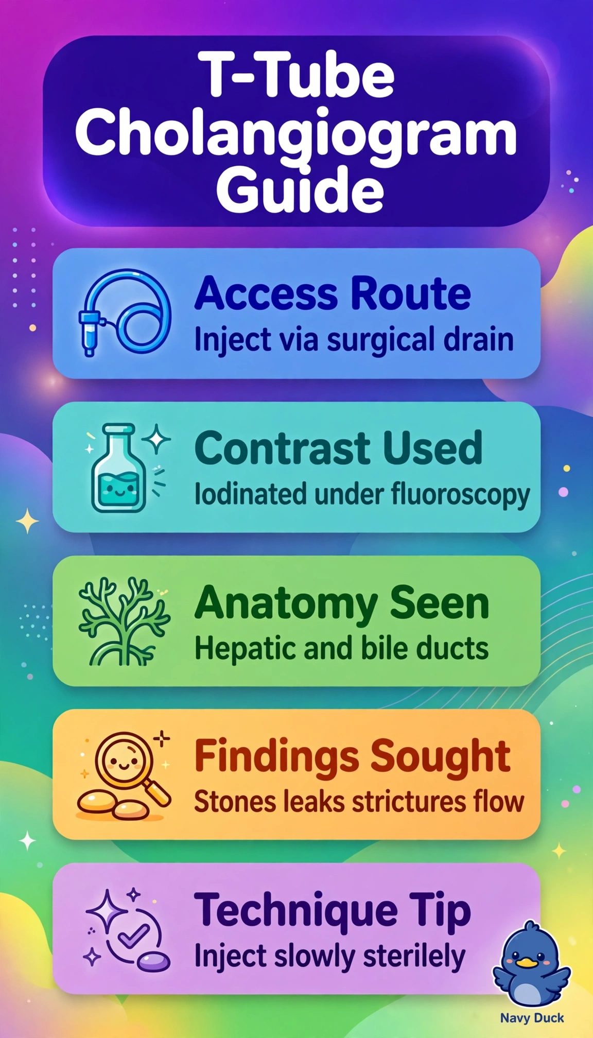
Surgical or T-tube cholangiography uses the indwelling biliary catheter placed at surgery to instill iodinated contrast under fluoroscopy, outlining the common hepatic and common bile ducts, showing filling defects from retained stones, strictures, or leaks, and confirming drainage into the duodenum. The barium enema approach fails because contrast enema evaluates the colon and cannot enter the biliary system, and barium is inappropriate for ductal injection, the ERCP approach fails because although ERCP also images bile ducts it requires gastroenterology endoscopy and ampullary cannulation rather than the surgical drain and is therapeutic as well as diagnostic, and the upper GI approach fails because it coats esophagus, stomach, and duodenum but does not opacify intrahepatic or extrahepatic ducts. Exam caveat: distinguish operative cholangiography during surgery from postoperative T-tube study and from ERCP by access route. Practical check: verify physician order, use sterile technique, inject slowly to avoid overdistention, and watch fluoroscopically for stones and free duodenal flow.