
A 45-year-old outpatient is scheduled for lumbar myelography for suspected spinal stenosis, the radiologist will perform a fluoroscopically guided lumbar puncture with iodinated contrast, and the radiographer is assigned to set up, assist sterilely, and monitor the patient in a small fluoroscopy suite. The consent and labs are verified and emergency equipment is present. Which radiographer assistance sequence should be used to support safe myelography and lumbar puncture technique?
Select an answer to reveal the explanation.
Short Explanation
Your job is keeping things clean, still, and watched closely. Sterile fluoroscopic assistance lets the doctor hit the sac safely on the first try.
Full Explanation
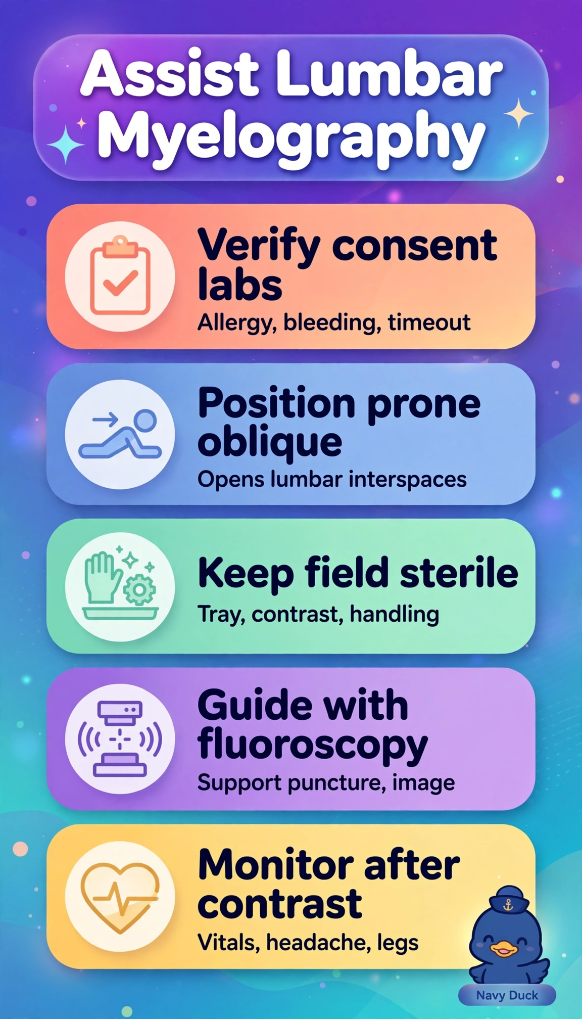
Safe lumbar myelography for suspected stenosis requires fluoroscopically guided prone-oblique positioning to open the interlaminar space and confirm needle entry and thecal sac flow, strict sterile setup and assistance for intrathecal puncture, and use of iodinated water-soluble contrast with post-injection monitoring of vitals and lower-extremity pain, weakness, numbness, or sphincter change suggesting irritation, bleeding, or compression. The gadolinium-based approach fails by contrast concept because intrathecal gadolinium belongs to distinct MR protocols with different dosing and safety considerations and cannot substitute for the specified iodinated fluoroscopic agent despite correct positioning and monitoring. The lateral decubitus bedside approach without fluoroscopy fails by guidance concept because although lateral decubitus is standard for bedside diagnostic puncture, this scenario explicitly requires fluoroscopic guidance with prone-oblique positioning to ensure accurate access and opacification. The clean-technique assembly fails by asepsis concept because although clean technique suffices for many general preparations, intrathecal injection demands maintenance of a sterile field to prevent meningitis and arachnoiditis. An exam caveat is to distinguish fluoroscopic iodinated myelography, often followed by CT, from MR myelography and to check whether monitoring includes both systemic vitals and focused neurologic leg assessment rather than vitals alone. A practical check is to verify identity, consent, allergies, and contrast name and concentration from the vial label with the radiologist, confirm sterile integrity and emergency equipment, and document pre- and post-procedure vitals with repeated leg checks before discharge and ordered follow-up imaging.