
A pediatric endocrinologist orders skeletal maturity assessment for a 5-year-old with early breast development and rapid height gain, and the clinic has no prior bone age images. The child is cooperative, the left hand is uninjured, and the parents ask why only a small body part is imaged. Which examination correctly completes bone age assessment in this patient?
Select an answer to reveal the explanation.
Short Explanation
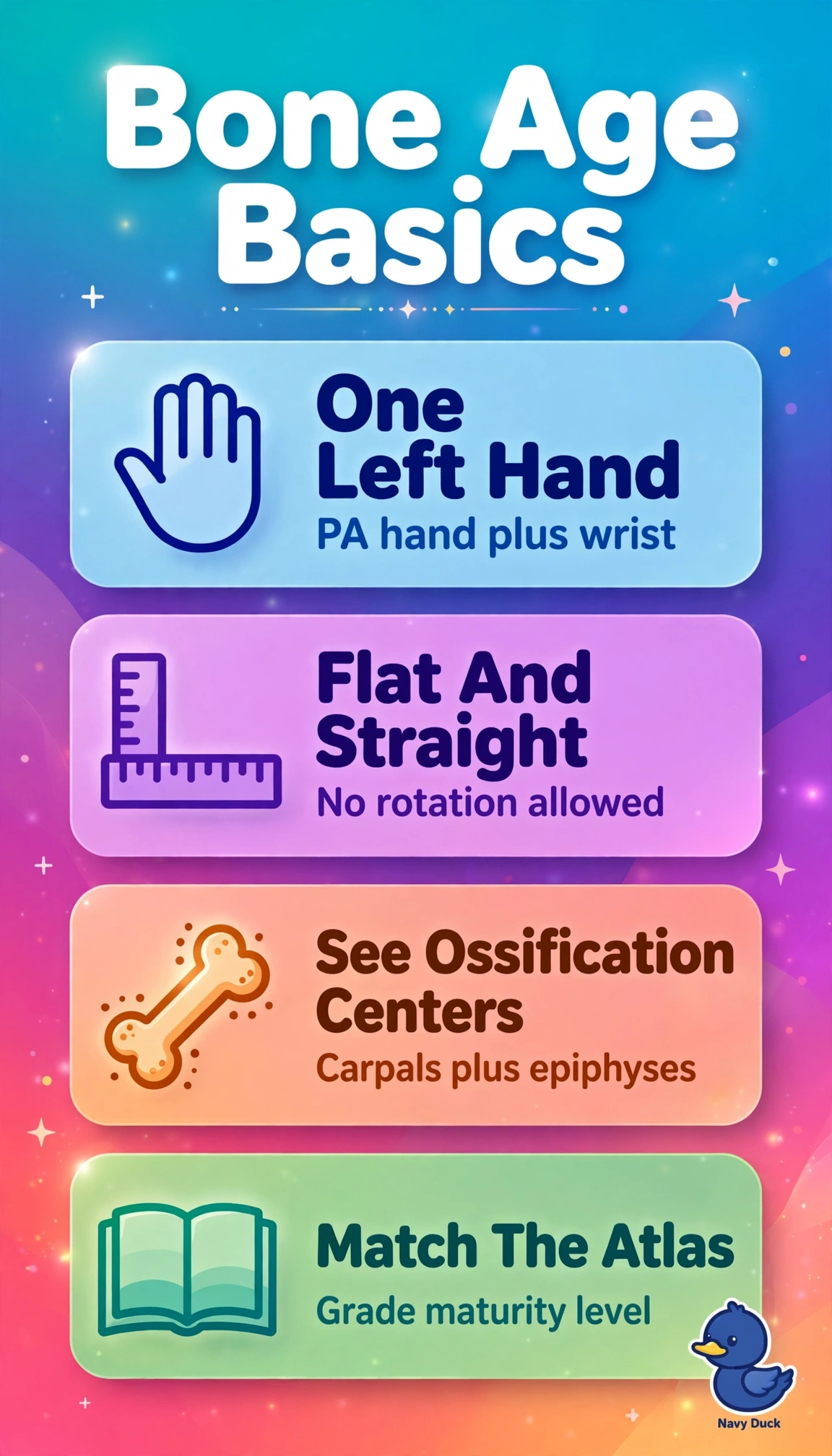
You’re comparing growth centers with a reference standard, not looking for fractures or change from an earlier study. A single left hand and wrist view shows the structures needed, and maturity is assessed from their pattern against standards.
Full Explanation
A single PA view of the left hand and wrist is the established baseline for bone age in a cooperative 5-year-old with suspected precocious puberty. Ossification of the carpals, metacarpals and phalanges correlates with validated Greulich-Pyle and Tanner-Whitehouse standards; the focused view also keeps radiation minimal, addressing the parents’ concern, and follows left-sided convention when the hand is uninjured. Using that view to assess fracture alignment and joint spaces is a trauma-focused approach: it does not systematically stage maturity against an atlas or provide the requested bone age. A left knee view is not interchangeable with hand-wrist standards for a mid-childhood precocity workup; although knee methods exist for specific infant or adolescent growth-remaining questions, it would not answer this indication. Measuring interval change from prior hand and wrist images requires a baseline, and none exists, so that approach would misrepresent a first assessment as follow-up. Exam caveat: bilateral, oblique or repeated views are not routine for uncomplicated bone age unless the left hand is injured, dysplastic or technically inadequate. Practical check: confirm true PA positioning, fingers extended together and slightly spread, neutral wrist, correct side marker and tight collimation to the hand and wrist; document that standards comparison was requested so the radiologist reports bone age with chronological correlation.