
A portable AP knee image shows the joint space closed, the patella displaced laterally, and one femoral condyle more distal than the other, with elongated tibial detail and increased density on one side. The technologist must decide if the error reflects positioning or tube-part-receptor alignment before repeating. Which evaluation of positioning accuracy and alignment best explains the image?
Select an answer to reveal the explanation.
Short Explanation
You're reading clues in the image to separate patient position from beam angle. Spotting rotation is like noticing a crooked photo frame on a wall. Straightening both patient and tube opens that joint.
Full Explanation
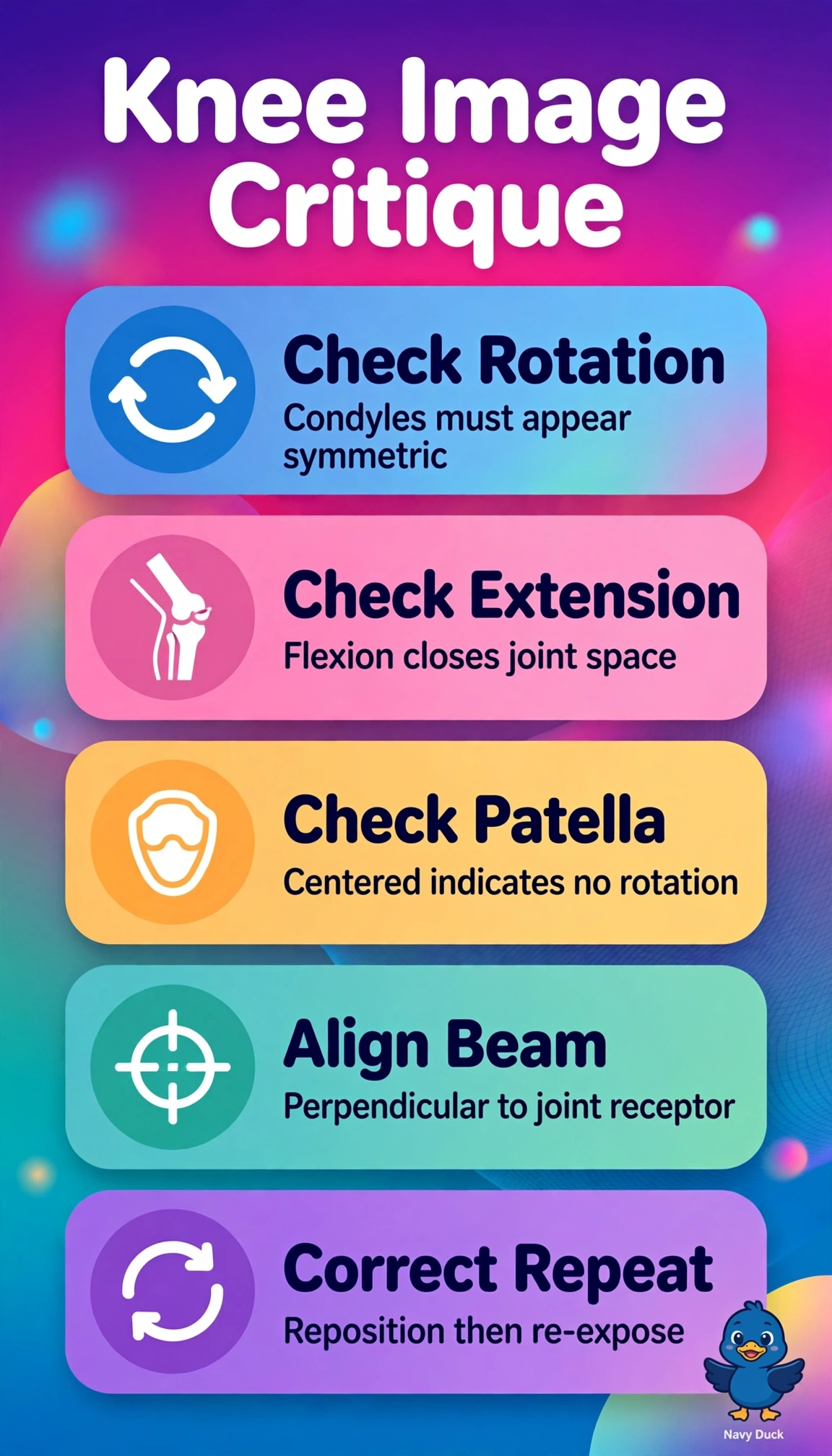
The correct evaluation integrates patient positioning with geometric alignment because lateral patellar displacement with one femoral condyle more distal signals rotation, closed joint space signals persistent flexion deformity without compensatory beam angulation parallel to the tibial plateau, and elongated tibial detail with asymmetric density signals distortion and differential attenuation from nonparallel tube, part, and receptor planes. Assessing insufficient cephalad angulation alone fails by beam-alignment concept because increasing angle may project through the joint but cannot recenter the patella or equalize condylar levels when the leg remains rotated and flexed, leaving the key positional signs unexplained. Assessing rotation and flexion while assuming parallel planes fails by distortion concept because elongation and unilateral increased density specifically indicate divergent-ray foreshortening and obliquity effects rather than pure anatomic malposition, so derotation without leveling the tube, knee, and receptor will still distort. Evaluating grid cutoff and heel effect alone fails by exposure-geometry concept because cutoff causes diffuse or peripheral underexposure with cutoff pattern and heel effect follows the anode-cathode axis, neither of which displaces the patella, staggers condyles, or closes the space. An exam caveat is that portable AP knees rarely match PA weight-bearing appearance, so test items expect recognition of combined rotation-flexion with malalignment rather than isolated exposure correction. A practical check is to center the patella, confirm symmetric condyles, palpate joint line, and align tube, part, and receptor parallel with the central ray to the joint before repeating.