
An adult ED patient has severe abdominal pain with suspected free intraperitoneal air. The patient cannot stand but can lie on the side with hips flexed. The surgeon needs air under the diaphragm or over the liver. Which abdomen positioning best demonstrates pneumoperitoneum when upright is impossible?
Select an answer to reveal the explanation.
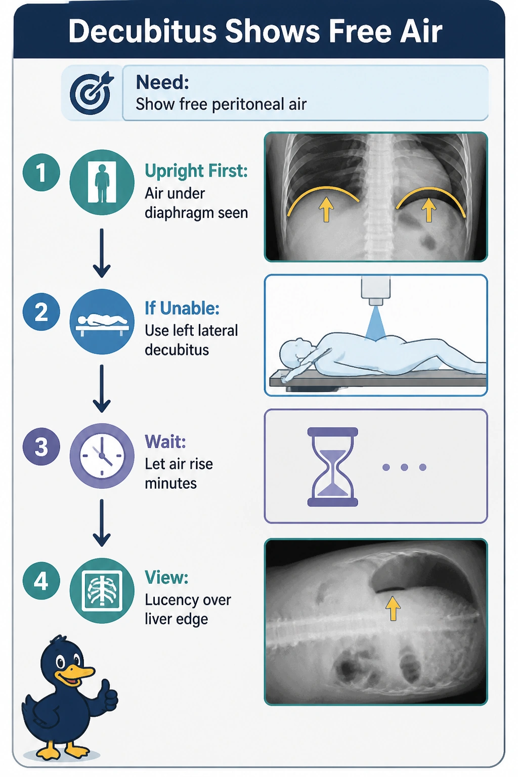
Short Explanation
You are letting the bubble float where you can see it. Rolling left side up sends free air to the liver edge like foam rising.
Full Explanation
Left lateral decubitus abdomen with horizontal beam is the preferred substitute when upright is impossible because with left side down the right flank and perihepatic space become nondependent, so free air rises and outlines the liver margin as a lucent band over the liver, directly satisfying the surgical need for extraluminal air. The erect upright abdomen view remains the classic standard for air under the diaphragm, yet it fails here on patient capability because the stem states the patient cannot stand but can lie on the side, making erect positioning unobtainable and unsafe despite its diagnostic quality. Right lateral decubitus abdomen uses the same decubitus principle and is routinely used for pneumoperitoneum, yet it fails on laterality and buoyancy because with right side down the liver is dependent and air migrates to the left upper quadrant around spleen and fundus, so no collection occurs over the liver. Left lateral decubitus assessment for fluid in the hepatorenal Morrison pouch is a common valid abdominal application, yet it fails on target physiology because fluid layers dependently while air rises to nondependence, so technique optimized for fluid misses the requested pneumoperitoneum. An exam caveat is that small pneumoperitoneum requires several minutes side-down for redistribution, a true horizontal ray, and inclusion of the entire up-side wall and liver edge, otherwise lung, colonic flexure or skin fold mimics air. A practical check is to verify markers for left-down right-up, keep hips flexed without rotation, center to include the liver dome, and confirm a sharp nondependent liver edge lucency before calling the study negative.