
A portable chest image on a ward workstation looks washed out with collapsed gray contrast, though exposure indicator and anatomy are acceptable. The surgeon wants better lung detail before rounds without repeating the exposure. Which postprocessing display control should the radiographer adjust first to improve visible contrast?
Select an answer to reveal the explanation.
Short Explanation
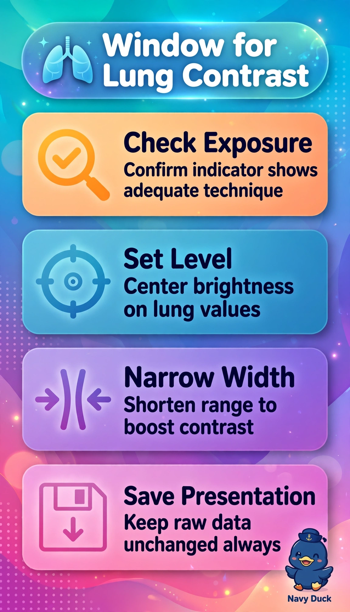
You do not need more dose for this fix. A quick tweak of window width and level rescues lung contrast in seconds.
Full Explanation
With an acceptable exposure indicator and complete anatomy, a washed-out portable chest reflects inappropriate display mapping rather than under- or overexposure, so narrowing window width around lung attenuation while setting level to aerated lung stretches subtle differences into diagnostic grayscale without repeat radiation. Increasing spatial frequency filtering enhances high-frequency edges and line sharpness but does not expand grayscale allocation, so contrast remains collapsed despite crisper borders. Applying dynamic range harmonization balances large-area density differences to display mediastinum and lung together, which addresses latitude and equalization rather than the narrow lung contrast needed for parenchymal detail. Reprocessing histogram segmentation recalculates field recognition and rescaling for brightness consistency, which cannot correct a display window error and is unwarranted when the indicator confirms correct detector exposure. Exam caveat: distinguish reversible presentation-state windowing on the ward workstation from permanent value-of-interest transformations or re-acquisition, as only the former meets the demand for immediate improvement without repeat exposure. Practical check: adjust width in small increments centered on lung, verify improved vascular and interstitial visibility without clipping mediastinal detail, then store the presentation for rounds.