
A school-age child with recurrent urinary infections is scheduled for voiding cystourethrography after a scout image, requiring bladder catheterization, iodinated contrast filling, and voiding views under fluoroscopy. Which cystourethrography protocol element best demonstrates bladder and urethral anatomy while maintaining sterile technique for this pediatric case?
Select an answer to reveal the explanation.
Short Explanation
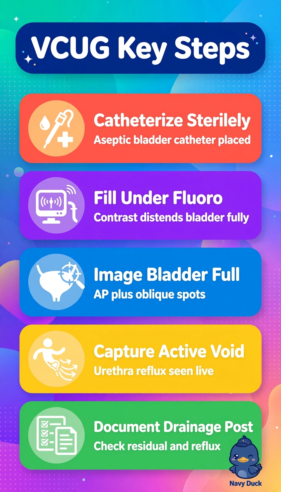
You reveal reflux and urethral detail by watching the bladder empty in real time. Sterile filling plus live voiding obliques catches the plumbing in action, like testing pipes under pressure, and voiding is the diagnostic moment.
Full Explanation
For recurrent infection in a school-age child, standard voiding cystourethrography starts with a scout image to assess bowel gas, calcification and bony landmarks, then fills the bladder with iodinated contrast through sterile catheterization under intermittent pulsed fluoroscopy. Imaging during active voiding in oblique projections elongates the urethra, separates bladder contours and best shows vesicoureteral reflux and urethral anatomy while limiting infection risk. Frontal imaging after voiding to assess residual volume is useful for retention evaluation, but is not the defining diagnostic view: after micturition, the distended urethra collapses, so valves, strictures and active reflux cannot be graded. Active voiding in frontal projection alone preserves the timing and sterile filling, but foreshortens the urethra and superimposes the bladder on distal ureters; obliques are needed to show the full urethral length and delineate reflux. Omitting the scout image while retaining oblique voiding views preserves dynamics and sterility but removes the baseline needed to recognize retained contrast, stool, calculi, enteric fistula clues or catheter malposition. Exam caveat: do not substitute nuclear cystography or intravenous urography when iodinated catheter filling and fluoroscopic urethral morphology are specified; nuclear imaging is excellent for low-dose reflux screening but lacks urethral spatial detail. Practical check: verify a sterile closed system, age-appropriate fill volume and tight collimation with ALARA pulsed fluoroscopy; document filling, full-bladder, active-voiding oblique and post-void images, along with the child’s tolerance.