
A 35-year-old with neck pain after whiplash needs routine cervical views plus open neural foramina, with no fracture on lateral and full head rotation possible. Which oblique cervical positioning best opens the lower neural foramina perpendicular to the image receptor?
Select an answer to reveal the explanation.
Short Explanation
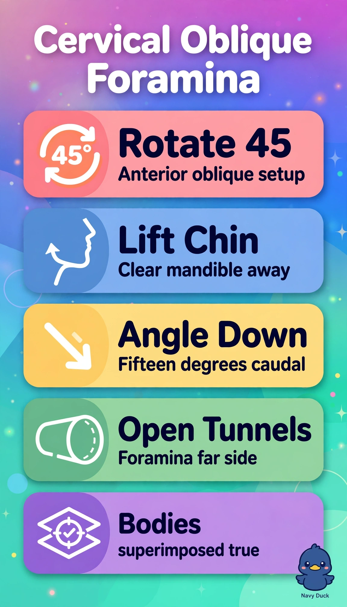
You’re lining up the lower foramina like open tunnels with 45-degree rotation, your chin extended, and a gentle caudal beam angle. This oblique view shows the foramina closest to the receptor clearly.
Full Explanation
For an anterior oblique cervical projection, rotate the body 45 degrees toward the side of interest, extend the chin to prevent mandibular overlap, and angle the beam 15 degrees caudally. This compensates for cervical lordosis and projects the intervertebral foramina closest to the receptor open and perpendicular to the beam, showing levels from C2-C3 through C7-T1, including the lower foramina. A 25-degree rotation with chin flexion under-rotates the patient; chin flexion closes the foramina and can superimpose the mandible. A perpendicular beam without caudal angulation projects the lordotic canals obliquely, making the foramina appear narrowed. A straight lateral with depressed shoulders shows alignment, disc spaces, and prevertebral soft tissues, but does not open the right and left foramina separately. Full hyperextension with a steep cephalic beam through the open mouth describes odontoid variants, not oblique foraminal anatomy, and risks pain after whiplash. Exam caveat: anterior obliques (RAO/LAO, caudal angle) show foramina closest to the receptor, whereas posterior obliques (RPO/LPO, cephalic angle) show those farthest from it. In practice, verify that rotation leaves the foramina open and clear of the pedicles, and collimate closely.