
A 54-year-old oncology patient in interventional radiology needs C-arm verification of a right basilic PICC tip at the cavoatrial junction, with no enteric tube in place and stable vitals. The physician asks the radiographer to provide pulsed imaging while advancing the catheter. Which C-arm imaging collaboration should the radiographer provide for this PICC line placement?
Select an answer to reveal the explanation.
Short Explanation
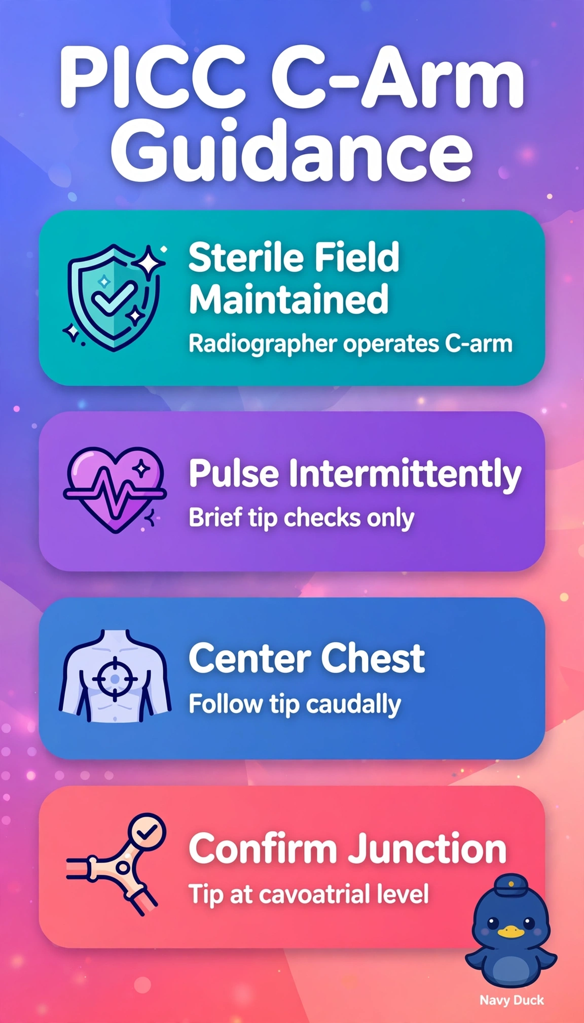
You're guiding a thread to the heart. Intermittent pulsed views light the path just enough without leaving the lights on.
Full Explanation
Intermittent pulsed C-arm guidance is correct because it balances real-time navigation of the right basilic PICC through the axillary, subclavian and brachiocephalic veins to the superior vena cava and cavoatrial junction with ALARA dose control in this stable oncology setting, allowing the radiographer to tap briefly during advancement, communicate direction and depth, and obtain collimated confirmation without excess exposure. Uninterrupted live screening during advancement fails by dose concept because continuous real-time exposure delivers substantially higher skin, scatter and staff dose and contradicts the explicit pulsed request, with no added steering benefit in a stable elective PICC when brief pulses suffice. Placing the tip just beyond the cavoatrial junction fails by target concept because the distal superior vena cava to cavoatrial junction is the accepted PICC terminus for reliable hemodilution and long-term oncology use, while atrial positioning risks arrhythmia, thrombosis, valvular irritation and mandated repositioning despite appearing secure. Verifying tip position only after advancement fails by timing concept because post-procedure documentation alone provides no steering through valves, tortuosity or venous variants, increasing passes, venous trauma and repeat imaging compared with active tracking. Exam caveat is to distinguish PICC acceptable zone from dialysis catheters or other central lines where deeper atrial placement is sometimes discussed, and not to apply enteric-tube or unstable-patient continuous monitoring logic here. Practical check is to pulse briefly during each advance, announce tip location relative to carina and right heart border, confirm free movement with respiration and flushing, then save the final cavoatrial junction image for the record.