
An older adult with a painful hip fracture, large body habitus, and limited mobility arrives by stretcher and cannot stand, rotate, or tolerate prolonged positioning for a trauma pelvis and lateral hip. Vital signs are stable but movement causes severe pain and the team needs diagnostic images quickly. Which procedural adaptation best addresses habitus, trauma, pathology, age, and limited mobility?
Select an answer to reveal the explanation.
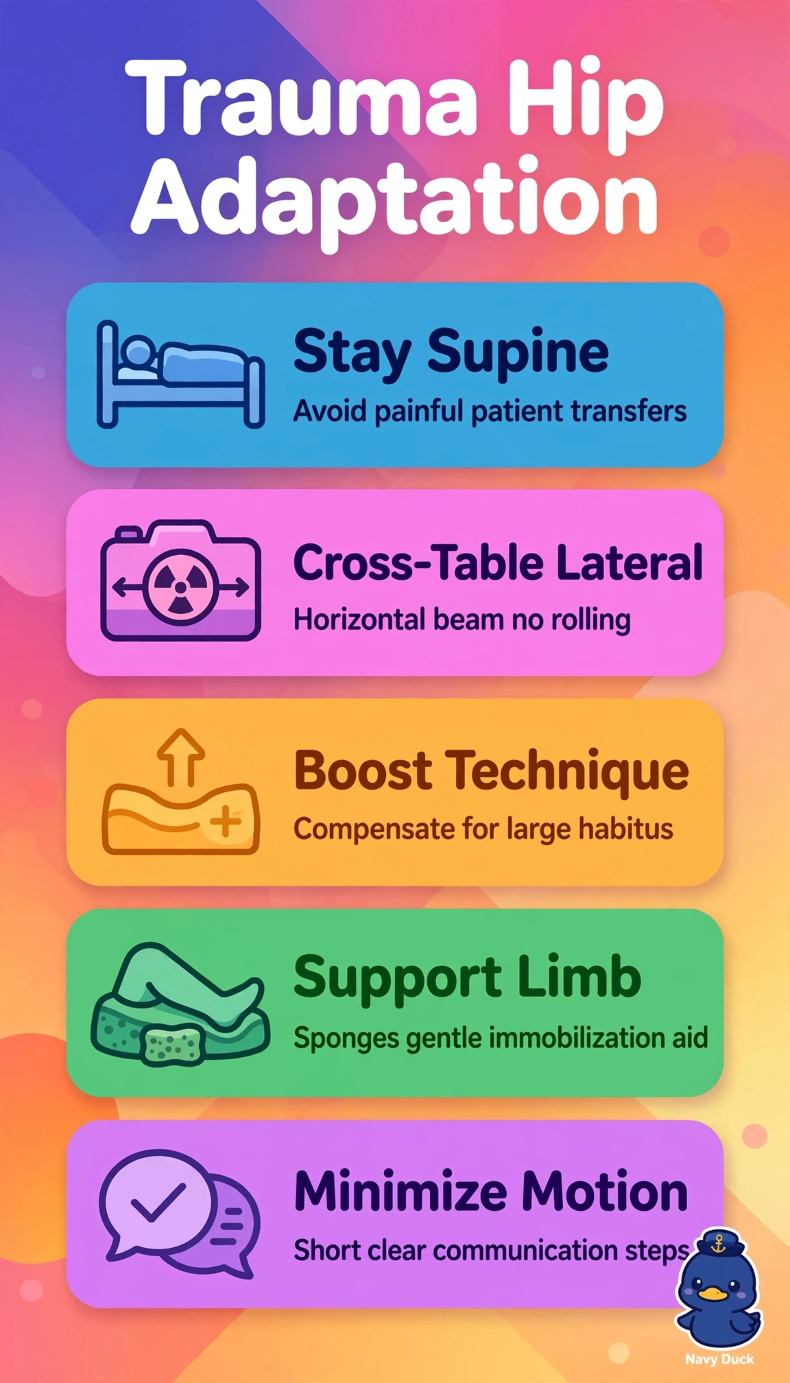
Short Explanation
You're bringing the exam to the patient instead of forcing the patient to the exam. Smart immobilization is like bracing a sore joint before moving it. Minimal motion means less pain and sharper images.
Full Explanation
The correct adaptation succeeds because it integrates all five constraints at once: remaining supine on the stretcher avoids standing, transfer, and rotation that would exceed limited mobility and provoke severe pain from the hip fracture in an older adult, the cross-table lateral provides the required orthogonal hip view without moving the injured limb, higher technique factors penetrate large body habitus to preserve density and contrast, and gentle immobilization support reduces motion while protecting fragile tissue. The standard-exposure variant fails on the habitus and image quality concept because dose restraint appropriate for average-size adults produces underexposure, noise, and nondiagnostic pelvis images through excess tissue, leading to repeats that increase total dose and delay diagnosis. The internal-rotation variant fails on trauma pathology principles because forced rotation for a routine true AP hip, although standard for intact anatomy, is contraindicated with suspected fracture as it risks displacement, neurovascular injury, and intolerable pain. The prolonged-obliques variant fails on tolerance and urgency because adding extended positioning for comprehensive views directly contradicts the inability to tolerate prolonged positioning and the need for rapid diagnostic images when movement causes severe pain. For exams, remember that trauma adaptation prioritizes obtaining two diagnostic orthogonal views safely over achieving ideal routine positioning, and increased technique for habitus is expected rather than penalized when penetration requires it. As a practical check, verify supine stretcher alignment with grid, select the large-habitus technique chart, place sponges and sandbags for support without pressure over the fracture, give brief clear breathing instructions, and reassess pain and neurovascular status immediately after the two exposures.