
A technologist performs an upright PA chest on a small adult using AEC with three ionization chambers, with the lungs centered to the middle chamber and the spine near the posterior chamber. The prior image was noisy from undertermination. Which AEC detector selection, alignment, and exposure adjustment best ensures proper AEC termination for this chest examination?
Select an answer to reveal the explanation.
Short Explanation
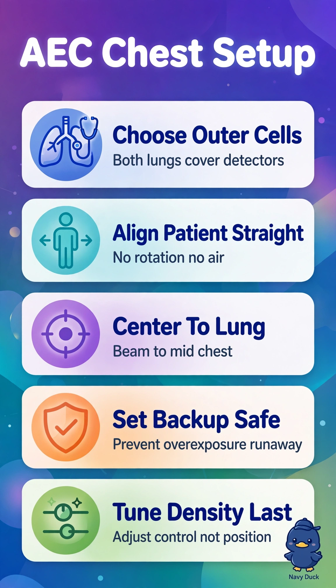
You get a clean chest when the measuring eyes actually see lung. Two outer cells over air-filled lung with straight anatomy lets the timer stop on time, like placing buckets under rain, and centering is everything.
Full Explanation
Proper AEC termination requires detectors receive transmitted radiation representative of the anatomy of interest, so for upright PA chest the two outer chambers must lie fully under aerated lung while avoiding dense spine and mediastinum and air outside the chest, with precise alignment and collimation to keep cells covered, plus a slightly increased backup time to allow completion in a small adult after undertermination without removing the safety ceiling. Lowering density control despite correct chambers and alignment fails on exposure logic because reduced density shortens integration to cutoff and repeats quantum mottle and noise, appropriate only when the prior image was overexposed rather than underterminated. Selecting all three chambers to average lung with mediastinum despite correct alignment and backup fails on detector selection because the central cells over high-attenuation spine delay cutoff and force lung overexposure while appearing balanced. Keeping correct outer chambers and backup but allowing relaxed centering that includes adjacent air fails on geometry because unattenuated photons reach the cells early and terminate prematurely, producing a light noisy image despite adequate backup. For examinations remember backup time never sets exposure, it only limits maximum time, so history of hitting backup in a small transmissive chest warrants a modest increase rather than a decrease. In practice confirm only outer cells are active, visually verify aerated lung fully covers them without spine or outside-air overlap, then check exposure index and whether backup terminated the exposure.