
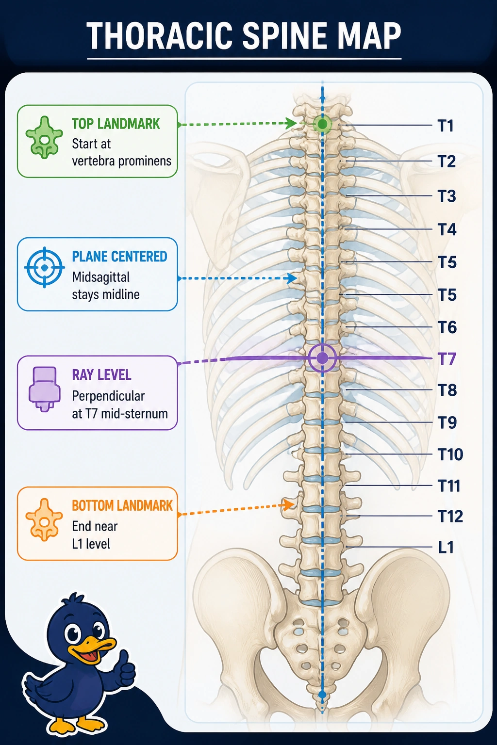
An ED trauma patient with mid-back pain after a fall needs a supine AP thoracic spine without moving the patient. The technologist must include all twelve thoracic vertebrae using surface landmarks, a true coronal alignment reference and a perpendicular beam. Which landmark, plane and central ray combination correctly includes the thoracic spine for this AP projection?
Select an answer to reveal the explanation.
Short Explanation
You're mapping chest bones to back bones without rolling the patient. A midline run from notch to tip centers T-spine, so T7 centering keeps all twelve vertebrae in view.
Full Explanation
The thoracic spine spans from the vertebra prominens (C7, just above T1) down to about L1 near the lower costal margin, lies in the midsagittal plane when supine, and is best profiled AP with the central ray perpendicular to the receptor at T7, which is mid-sternum, midway between the jugular notch and xiphoid, to place the long column centrally with minimal distortion. That landmark, plane and ray combination includes T1 through T12 in a trauma-safe supine position. Iliac crest to pubic symphysis with midcoronal centering and angulation toward the femoral heads describes an AP pelvis or hip, missing the thorax entirely. Vertebral prominens to iliac crest with L3 centering describes a lumbar spine length, excluding the upper thorax. Sternal angle to umbilicus with midaxillary centering and T12 centering shifts inferior and lateral, clipping upper thoracic vertebrae and introducing obliquity. Exam caveat: planes describe patient orientation, landmarks set collimation borders, and ray level sets centering — all three must agree. Practical check: palpate notch and xiphoid, center midsagittal to the table, and set the perpendicular ray to mid-sternum.