
A new DR room shows fine grainy stripes and slight shading across flat phantom images, plus posterized gray steps in soft tissue on clinical images. Service suspects digitization and calibration before the histogram is even built. Which raw-data preprocessing step should be checked first to correct detector signal uniformity?
Select an answer to reveal the explanation.
Short Explanation
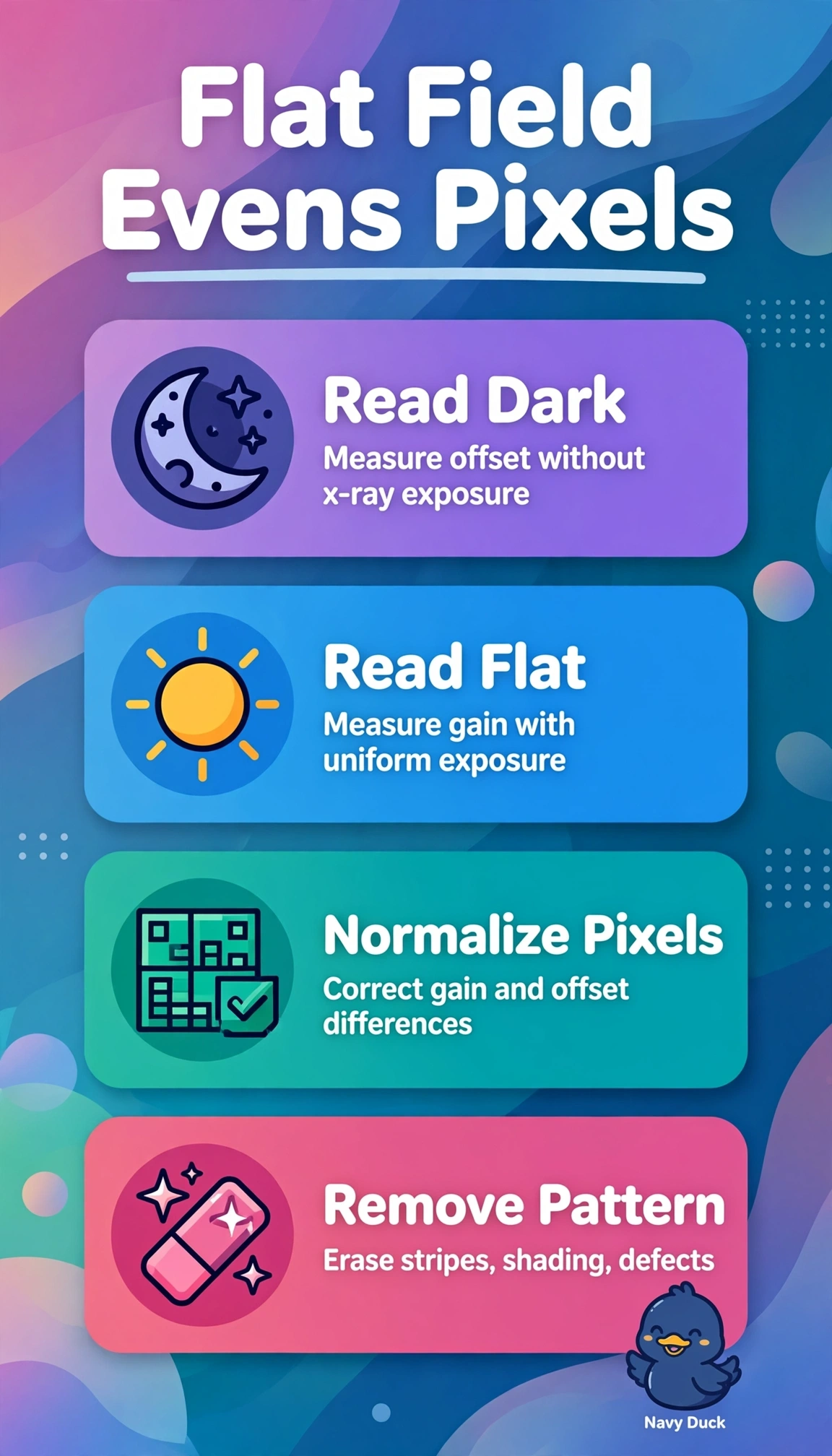
You’re looking before the image is polished. Even shading and stripes point to flat-fielding, which corrects detector uniformity.
Full Explanation
Flat-fielding is the appropriate first check because fine stripes and broad shading on uniform phantom exposures indicate spatial gain and offset variation across detector elements. Flat-field correction uses uniform calibration exposures to apply per-pixel gain normalization and offset correction, making the raw panel response more uniform before further processing; this can also help preserve subtle soft-tissue gradation when detector nonuniformity disrupts low-contrast detail. Dark-offset subtraction is a valid preprocessing step, but it removes only additive electronic background measured without X-rays; it does not compensate for X-ray-dependent sensitivity differences that cause shading and striping under exposure. Defective-pixel interpolation replaces isolated dead or hot pixels, or defective lines, with values derived from neighboring pixels, but does not correct broader pixel-to-pixel gain variation or shading. Temporal lag and ghosting correction addresses residual signal carried over from previous exposures, not stable spatial nonuniformity across phantom images. An exam caveat is that wording about correction before histogram analysis points to detector-level calibration, rather than later image display processing. A practical check is to acquire fresh flat phantom images after dark-offset and flat-field recalibration according to service protocol, then confirm that the images show uniform variance without stripes or shading before assessing clinical soft-tissue contrast.