
A 62-year-old outpatient with large body habitus and a thick plaster lower-leg cast returns for AP and lateral ankle follow-up, has moderate pitting edema beneath the cast, and prior images appear pale with poor bony detail despite standard adult ankle technique. Which technique adaptation best restores bony detail through cast and tissue?
Select an answer to reveal the explanation.
Short Explanation
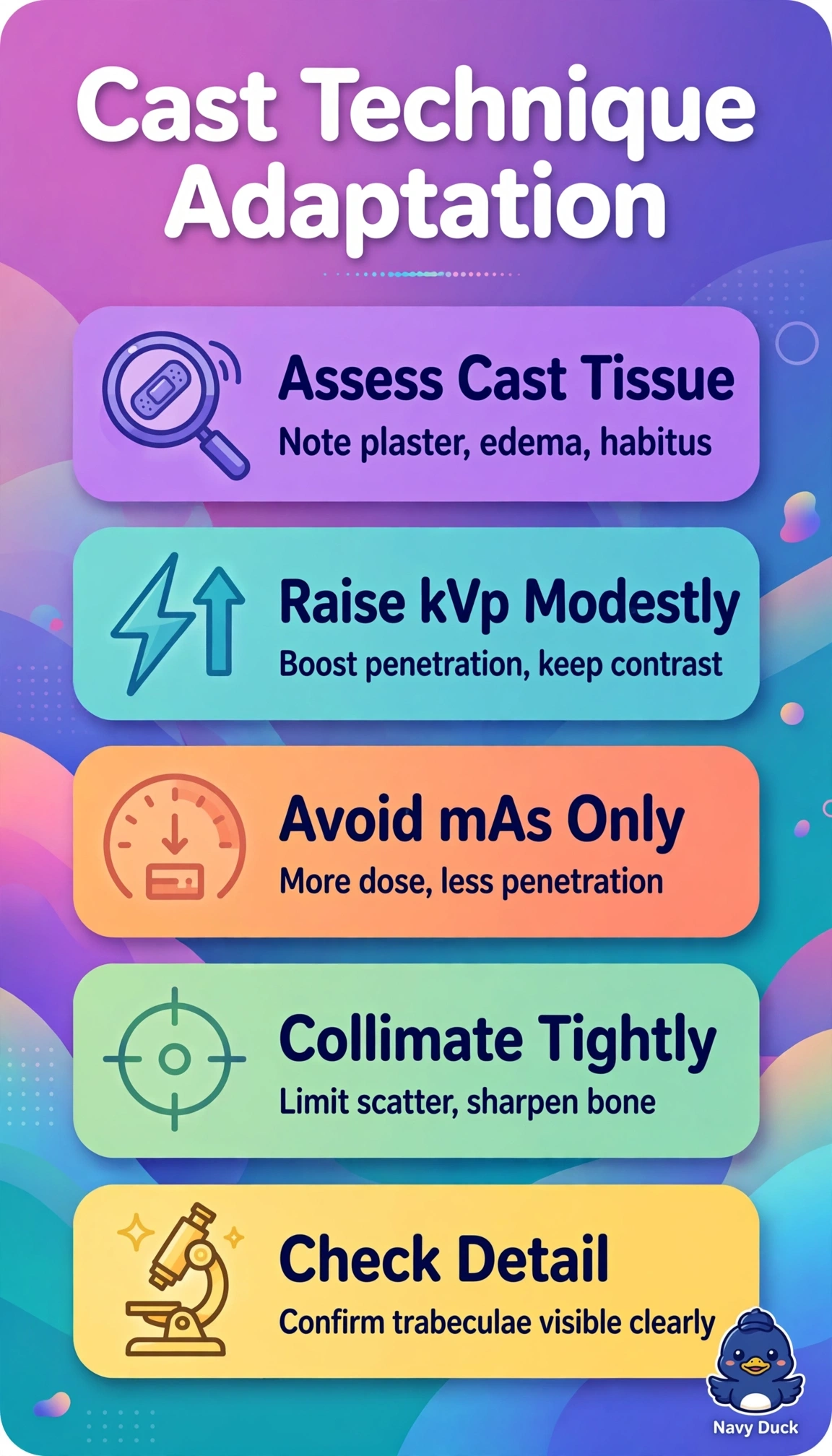
You need more beam penetration to get through the plaster and extra tissue. A modest kVp boost helps reach the receptor while keeping usable contrast, like turning up water pressure to rinse thick mud.
Full Explanation
Plaster, edema, and large body habitus all increase beam attenuation, so moderately increasing kVp raises photon energy and penetration, helping enough signal reach the receptor to show trabecular bone through the cast while preserving an acceptable scale of contrast. Decreasing kVp substantially fails because, although lower energy increases photoelectric interaction and subject contrast in theory, it sharply reduces penetration and can leave the image even paler and mottled from photon starvation. Keeping standard adult technique unchanged fails to adapt to the added cast material and tissue thickness; increased attenuation can produce underexposed, nondiagnostic images that may require a repeat. Raising mAs by only 10 percent without changing kVp also fails to compensate for substantial attenuation from thick plaster, edema, and large habitus, leaving inadequate receptor exposure and mottled detail. For the exam, associate plaster and large habitus with a moderate rather than drastic kVp increase. In practice, verify the cast type and thickness, adjust in small increments, collimate tightly, and review bony detail before considering a repeat. A practical caveat is to preserve usable contrast rather than increase kVp so much that the image’s contrast scale becomes unsuitable.