
A repeat abdomen series shows acceptable contrast but excessive noise at 70 kVp and low mAs without AEC, and the radiologist requests lower patient dose while preserving diagnostic quality for an average adult. The grid, SID, and collimation will remain constant for the repeat exposure. Which exposure factor change best balances image quality and dose effects?
Select an answer to reveal the explanation.
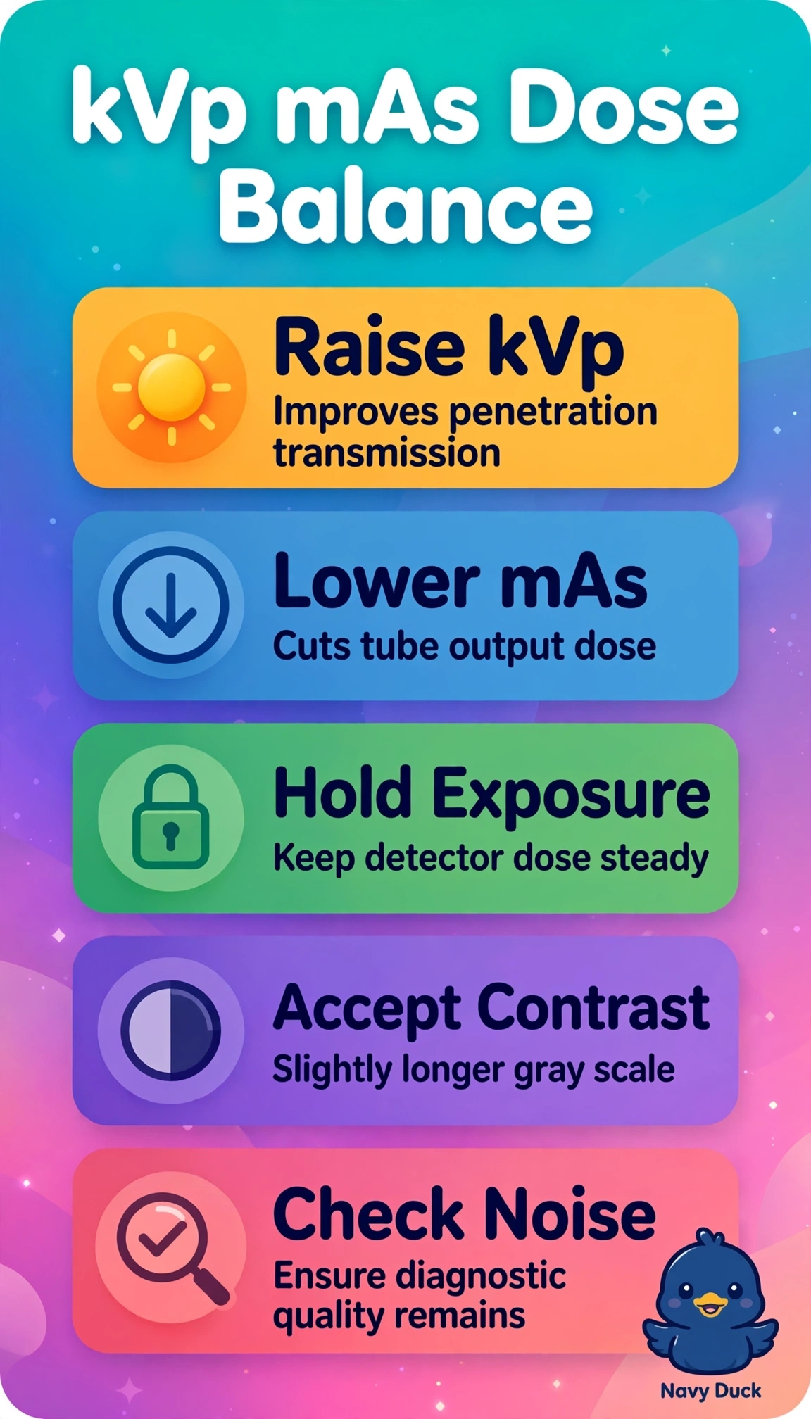
Short Explanation
You're trading a little contrast for a lot of dose savings when you use the right balance. Raising kVp works like opening a wider faucet to let more penetrating photons through. Lower mAs then keeps the detector happy with less skin dose.
Full Explanation
The correct change works because 70 kVp is relatively low penetration for an average adult abdomen, leaving acceptable contrast but photon-starved quantum noise, so a moderate kVp increase improves beam transmission and detector signal per unit dose while a proportional mAs reduction keeps detector exposure stable, thereby lowering entrance dose while preserving diagnostic noise and contrast with grid, SID and collimation unchanged. Raising mAs alone at constant kVp is a standard method to smooth quantum mottle and would address the stated noise, but it directly increases tube output and patient dose and therefore violates the explicit request for lower dose. Lowering mAs alone at constant kVp is a standard dose-saving method when image quality is ample, but with noise already excessive it further reduces photon fluence to the detector and degrades signal-to-noise and diagnostic acceptability despite maintaining contrast. Lowering kVp with compensatory higher mAs is a standard method to enhance subject contrast in small parts or low-contrast studies, but here contrast is already acceptable and the adult abdomen requires penetration, so reduced energy increases photoelectric absorption and required mAs, raising dose and potentially worsening noise. An exam caveat is to treat the 15 percent kVp rule as an approximate exposure-maintenance guide rather than exact dose equivalence and avoid large kVp swings that would flatten needed contrast or exceed latitude. A practical check is to compare exposure index, deviation index, visual noise and DAP against the prior image to confirm stable detector exposure with reduced dose.