
A technologist images three outpatients in sequence: a child’s forearm in a fiberglass cast, an adult with advanced osteoporotic wrist, and a large-BMI adult knee, all with prior overexposed repeats. Which technique adaptation best balances diagnostic density while respecting pathology, age, and cast attenuation?
Select an answer to reveal the explanation.
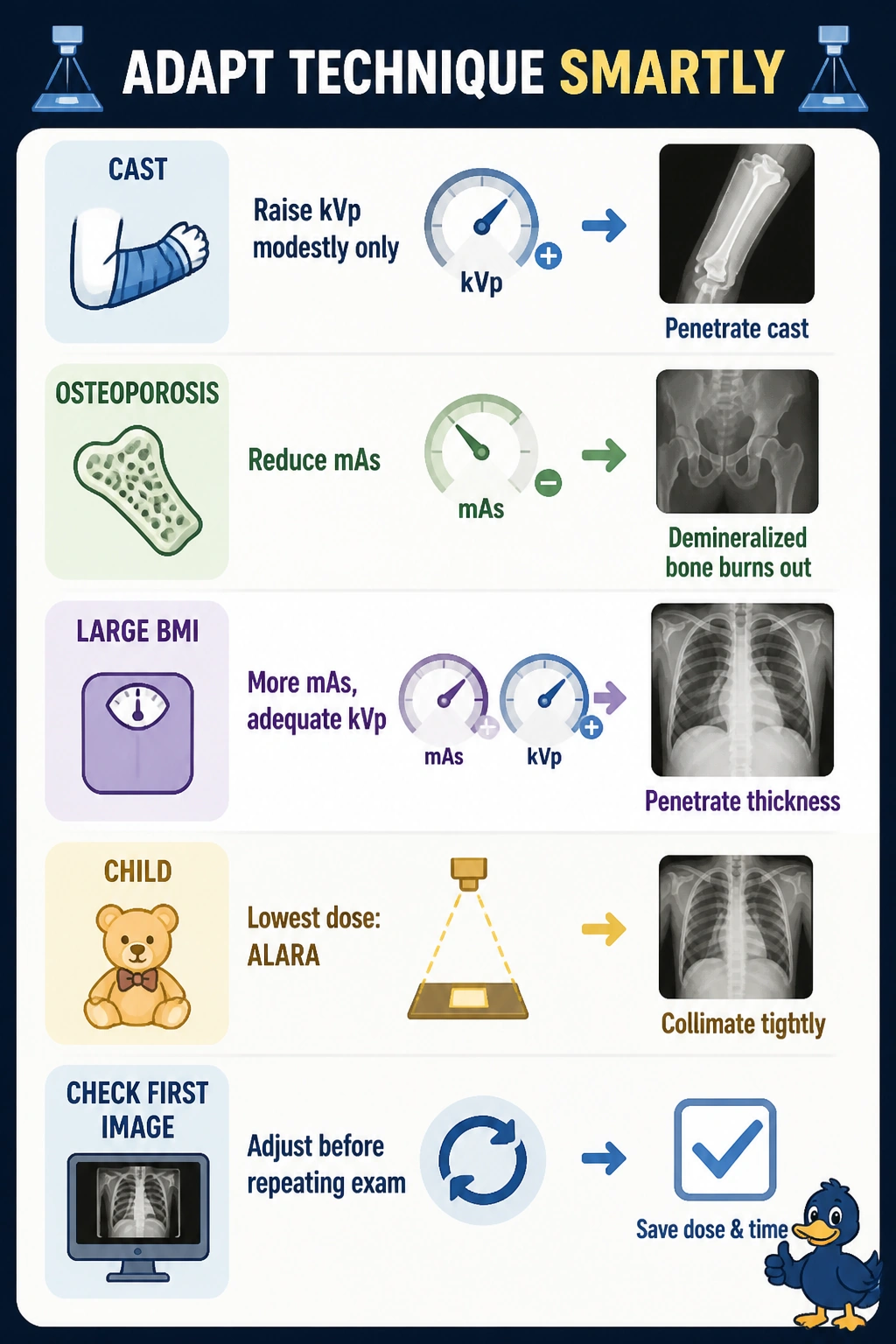
Short Explanation
You're juggling three very different patients, so you'll tweak your technique like adjusting headlights for fog, glare, or a dark road. Give the cast a modest kVp bump to punch through, lighten mAs for that fragile osteoporotic wrist, and boost both for the larger knee to keep your density diagnostic without repeats.
Full Explanation
Casts attenuate the beam and typically require a modest kVp increase to maintain penetration without excessive mAs and dose, osteoporotic or demineralized bone has reduced attenuation and needs reduced mAs to avoid overexposure and loss of trabecular detail, and large BMI increases attenuation and scatter requiring increased mAs with adequate kVp plus grid or beam restriction as protocol dictates, while pediatric patients warrant the lowest exposure that remains diagnostic. Decreasing factors for a cast produces light, noisy images, substantially increasing mAs for osteoporotic bone blows out contrast, and decreasing kVp for large habitus reduces penetration and increases mottle. Using identical adult factors ignores age, pathology, and size, raising dose to the child and osteoporotic patient while underexposing the large knee. Markedly increasing mAs with low kVp for casts adds dose without penetration, high kVp for osteoporosis washes out delicate bone detail, and cutting mAs for large BMI guarantees quantum mottle. The exam caveat is to adapt for病ology and habitus while honoring ALARA and pediatric sensitivity, and the practical check is to confirm cast material and thickness, assess bone density on prior images, measure part thickness, and evaluate the first image before repeating.