
A quality-assurance audit flags rising repeat rates and creeping exposure indicators on DR wrist images, with detectors showing wide latitude masking overexposure. The team must reduce dose while maintaining diagnostic quality and documentation. Which strategy best addresses detector efficiency, latitude, and dose tracking for this audit?
Select an answer to reveal the explanation.
Short Explanation
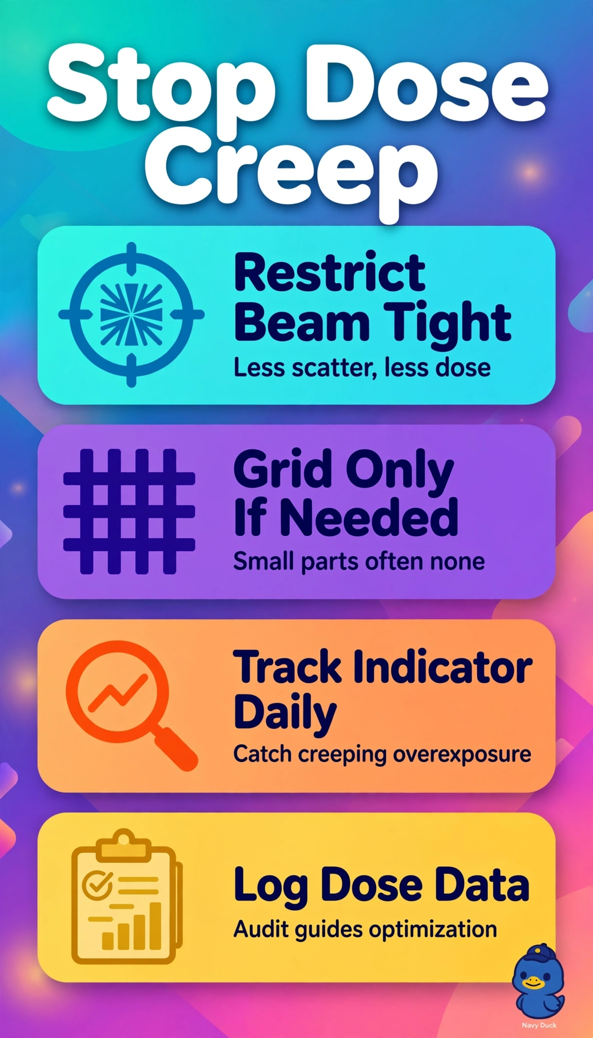
You can curb dose creep while keeping wrist images useful and the audit trail complete. Exposure indicators reveal detector dose that wide DR latitude can hide.
Full Explanation
Optimizing collimation and grid selection, monitoring exposure indicators, and logging dose indices best addresses detector efficiency, latitude, and dose tracking. Collimation limits irradiated tissue and scatter, while appropriate grid selection balances detail against unnecessary dose; indicator review can reveal upward dose creep hidden by wide DR latitude, and dose-index logs provide documentation for audit. Standardizing wrist positioning and exposure technique, monitoring indicators, and logging dose indices supports repeat reduction and tracking, but omits collimation and grid optimization, leaving field size and scatter uncontrolled and missing the efficiency requirement. Optimizing collimation and grid selection, monitoring indicators, and coaching staff on visual image quality addresses dose control but omits dose-index logging, so it leaves no auditable record. Optimizing collimation and grid selection, assessing sharpness and positioning, and logging dose indices supports image assessment and documentation but substitutes visual assessment for indicator monitoring, allowing latitude to mask overexposure. Exam caveat: acceptable brightness does not prove acceptable detector exposure in DR, because processing can rescale a wide input range. Practical check: compare each wrist exposure indicator with the manufacturer’s target range and correlate logged deviations with repeat analysis before changing technique.