
A 28-year-old warehouse worker fell onto the shoulder during a morning shift and has point tenderness over the acromioclavicular region without deformity suggesting dislocation. The patient can stand upright and lift light weights symmetrically, and the physician suspects a low-grade separation. Which AC joint projection protocol best demonstrates joint separation in this patient?
Select an answer to reveal the explanation.
Short Explanation
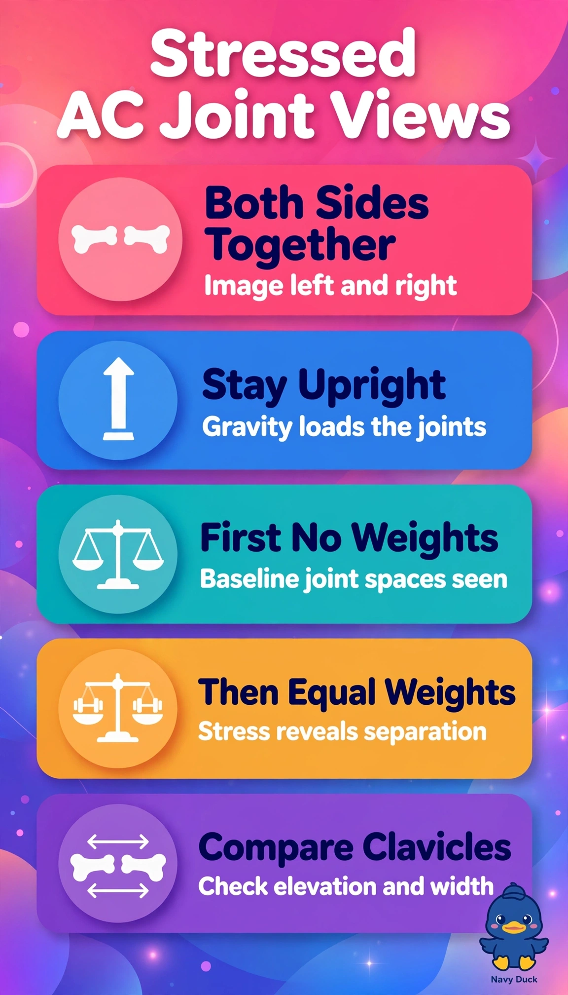
You’re comparing tiny joint spaces side to side, so symmetry is everything here. Bilateral upright views without weights and then with equal weights can make even subtle widening stand out, and symmetry is your secret weapon for AC injuries.
Full Explanation
Low-grade acromioclavicular separation is assessed by comparing clavicle elevation and joint width under physiologic stress. Bilateral upright AP AC views, first without weights and then with equal weights, provide matching sides and load the supporting ligaments while gravity distracts the shoulder girdle. A Grashey AP oblique profiles the glenohumeral joint for dislocation or arthritis, but unilateral rotation prevents side-to-side comparison and does not stress the coracoclavicular ligaments. An upright scapular Y lateral shows humeral head position relative to the glenoid for dislocation and scapular fractures, but superimposes the AC joint and provides no loaded comparison. A supine axillary Lawrence projection profiles the glenohumeral relationship and coracoid region for dislocation, but requires abduction that may be painful, images only one side, and provides no gravity stress. Exam caveat: a routine shoulder projection, including a Grashey view, does not replace dedicated stressed bilateral AC views when separation is suspected. Practical check: confirm upright posture, no rotation, equal weights held just off the floor, and marker placement before exposure.