
A community orthopedic clinic images AP pelvis on adults from 110 to 250 pounds, wants consistent trabecular contrast for arthritis grading, currently bumps kVp for larger patients and sees washed-out joints, and repeat rates are rising. Which technique-chart strategy best maintains consistent density and contrast across body habitus for this pelvis workload?
Select an answer to reveal the explanation.
Short Explanation
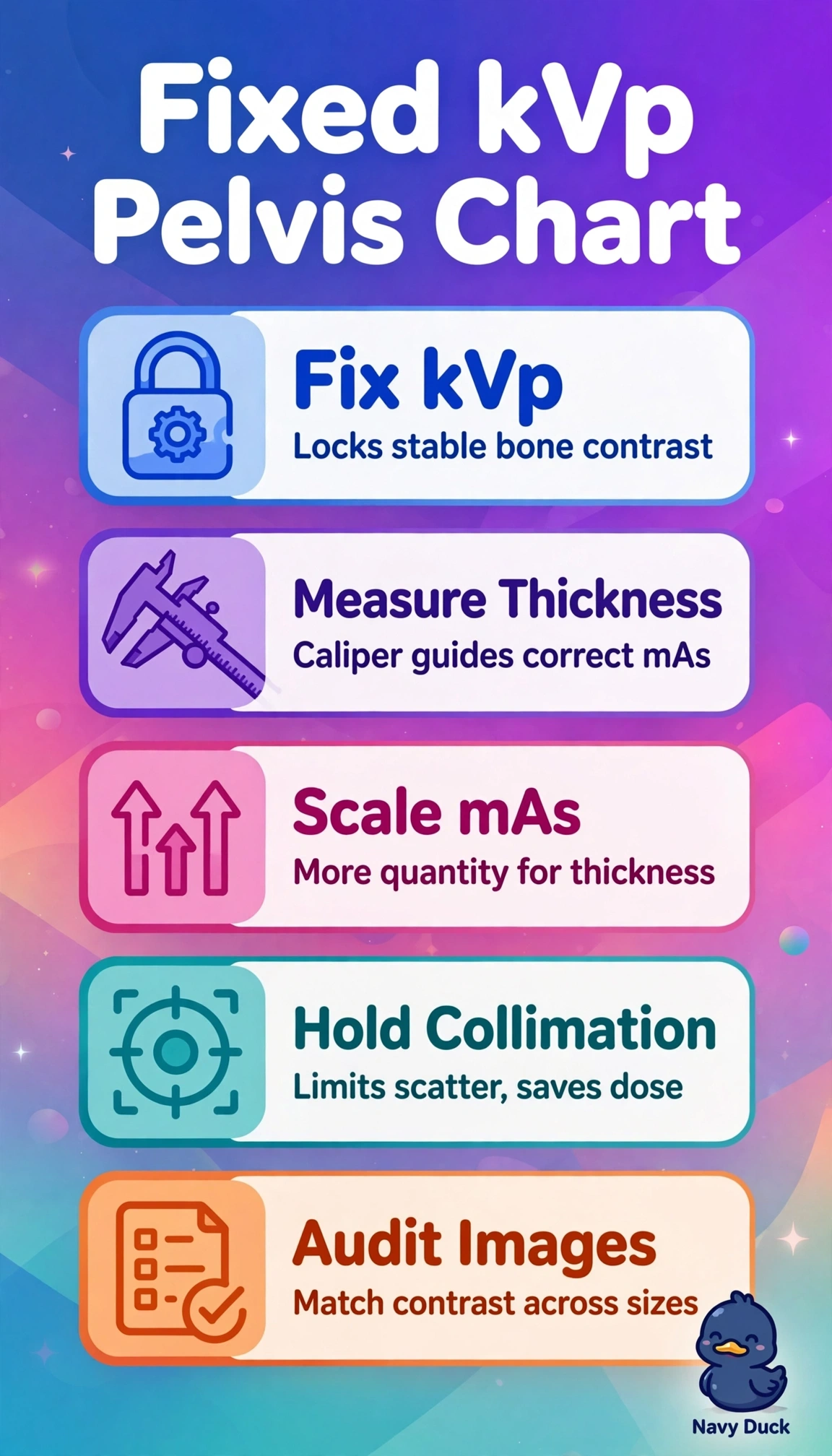
Your washed-out joints come from chasing penetration with kVp, which flattens contrast. A fixed-kVp technique keeps bone contrast steady while mAs handles thickness—like keeping the same filter and adding light.
Full Explanation
A fixed-kVp chart holds photon energy and beam quality steady, helping maintain subject contrast in the pelvis for grading joint spaces and trabeculae across body sizes. You then control density by scaling mAs to thickness, often measured with calipers; this increases photon quantity without changing beam quality. Raising kVp by 2 per centimeter while holding mAs constant fails by the contrast concept: higher kVp increases Compton scatter and reduces differential attenuation, washing out large-pelvis images even when density is adequate. Holding mAs constant at a fixed kVp preserves contrast but fails by the density concept: large patients may be underpenetrated and small patients overexposed, producing noisy or saturated images. Sharply raising both kVp and mAs for large patients fails by dose and contrast concepts: it overcompensates quantity while higher energy degrades contrast, increasing patient dose without diagnostic gain. Exam caveat: anatomically programmed radiography is consistent only when programmed around fixed-kVp logic and regularly calibrated to thickness. Practical check: caliper the pelvis, select the charted mAs for that thickness at the fixed kVp, and review whether contrast is matched before adjusting the programming.