
A hand clinic needs crisp detail of a scaphoid fracture line without motion blur, using a small focal spot, short exposure, and proper tube alignment. The anatomy is thin and requires high spatial resolution with uniform density. Which technical selection best optimizes recorded detail while managing the anode heel effect?
Select an answer to reveal the explanation.
Short Explanation
You squeezed every line pair out of that wrist. Cathode placement bathes thin anatomy in the stronger beam side.
Full Explanation
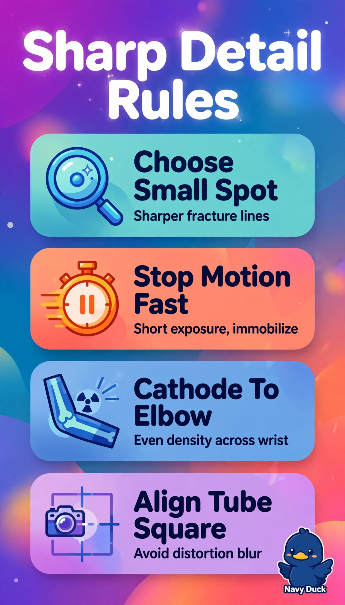
The correct selection succeeds because thin scaphoid anatomy demands high spatial resolution from a small effective focal spot that limits geometric unsharpness while short exposure with firm immobilization freezes subtle trabecular detail, and orienting the more intense cathode side toward the thicker proximal forearm near the elbow with the lower-intensity anode toward the thinner fingers compensates for differential absorption from the anode heel effect to produce uniform density, with close collimation further reducing scatter and improving contrast. A reversed cathode-anode orientation using a small focal spot and close collimation fails by heel effect principle because placing higher intensity distally over thin fingers while placing lower intensity proximally over thicker wrist creates nonuniform density and risks underexposure proximally despite excellent sharpness. A technique that preserves correct orientation and small focal spot but substitutes extended exposure time fails by motion control principle because even submillimeter movement during a longer exposure blurs fine fracture lucency despite adequate density and resolution. A technique that preserves correct orientation and collimation but substitutes a large focal spot fails by resolution principle because larger effective source size increases penumbra and geometric unsharpness that obscures hairline fracture margins despite better heat capacity and shorter tube loading. Exam caveat is to distinguish heel effect uniformity from magnification or grid alignment, as angling or SID changes do not compensate for intensity variation along the cathode-anode axis. Practical check is to verify cathode location marked on tube housing, confirm thickness gradient from elbow to fingers before positioning, and review a test image for even density and crisp cortical margins without doubling.