
A 16-year-old with suspected adolescent idiopathic scoliosis needs an initial standing curvature assessment, and her 68-year-old grandmother needs sacroiliac joint pain evaluation after a fall without fracture on lumbar views. Which projection pairing best demonstrates spinal curvature and SI joint anatomy for these two patients?
Select an answer to reveal the explanation.
Short Explanation
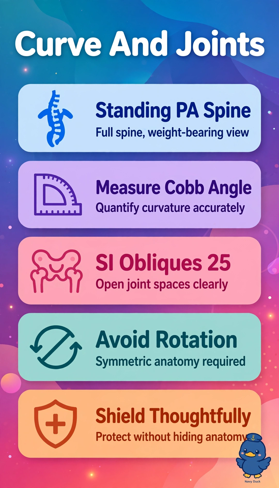
You've got two different jobs here — you'll check her curve with a standing PA view, like sizing up a tower while it's bearing weight so you see the real lean. For grandma, you'll add steep posterior obliques to swing those tucked-away joints open into view.
Full Explanation
A standing posteroanterior scoliosis series includes the full spine from cervical to sacrum to measure Cobb angles under physiologic load while PA positioning reduces breast and thyroid dose compared with AP, and bilateral posterior obliques at about 25 degrees open the sacroiliac joints by projecting the joint spaces en face for erosion, sclerosis, or diastasis evaluation. A supine AP thoracolumbar view unloads the curve, underestimates severity, limits Cobb measurement, and a straight AP pelvis superimposes the ilium and sacrum over the SI joints without opening them. A single lateral scoliosis view profiles kyphosis but cannot measure lateral curvature, and 45-degree anterior obliques misalign the SI joint plane and flexion degrades joint sharpness. Recumbent rotated obliques distort curvature assessment and a lateral sacrum alone superimposes the SI joints rather than separating them. The exam caveat is to use PA erect for scoliosis when possible with gonadal shielding that does not obscure anatomy, and the practical check is to ensure no rotation, include iliac crests and hips as protocol requires, and verify SI joint spaces are open and symmetric.