
A 25-year-old athlete has a mediolateral left knee projection for ligament injury, the image shows the femoral condyles, patella, and joint space, but rotation and tube-part-receptor alignment are uncertain. Which image evaluation criterion confirms accurate positioning with proper alignment for this mediolateral knee?
Select an answer to reveal the explanation.
Short Explanation
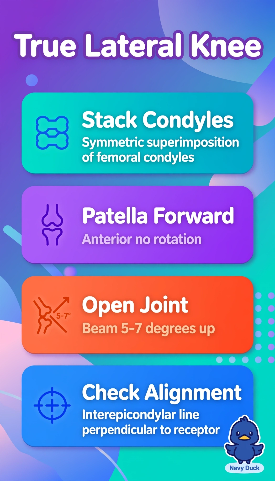
You're checking a true side view, not a slanted peek, so symmetry tells the tale. Superimposed condyles stack like coins when rotation and beam angle are spot-on.
Full Explanation
Symmetric superimposed condyles with anterior patella and open joint space is correct because a true mediolateral knee requires the femoral epicondyles directly superimposed indicating no rotation, the central ray directed 5-7 degrees cephalad to place the joint space open, and the patella anterior without magnification distortion confirming lateral tube-part-receptor alignment. The fibular head separated approach fails because that describes an AP or oblique knee projection criterion where fibular head position gauges rotation, not a lateral view. The patella centered in groove approach fails because that describes a sunrise or Merchant patellar view evaluation for patellofemoral congruence, not mediolateral alignment. The femoral neck elongated approach fails because that describes proximal femur and hip projection assessment, anatomically distant and irrelevant to knee joint positioning. Exam caveat is to memorize one hallmark per projection: laterals need superimposition, AP needs symmetry, sunrise needs centered patella. Practical check is to flex knee 20-30 degrees, place epicondyles perpendicular to receptor, angle beam slightly cephalad, and repeat only if condyles differ by more than half centimeter.